 |
| About Bioline | All Journals | Testimonials | Membership | News |
|
||||||
|
||||||
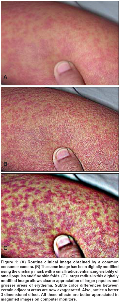
Indian Journal of Dermatology, Venereology and Leprology, Vol. 75, No. 2, March-April, 2009, pp. 191-193 Letter to the Editor Enhancing digital images using unsharp-mask Taneja Atul Apollo Gleneagles Hospitals, Kolkata-700 054 Code Number: dv09059 Sir, The practice of dermatology requires clear visualization and delineation of particular regions of interest on the skin. During clinical examination, dermatologists attempt to visually isolate and distinguish areas of interest from the surrounding skin by changing angles of observation, using oblique lighting, low illumination, the Wood′s lamp and other commercial equipment. Digital images of clinical lesions are now-a-days routinely obtained and these images can be manipulated on any ordinary computer in a variety of ways to similarly enhance the regions of interest. One such simple manipulation used for sharpening edges is called the unsharp-mask. This feature is inbuilt within a variety of photoediting software like Adobe Photoshop R , which enhances contrast between any two or more adjacent pixels. Any properly exposed, well-balanced, digital image of the skin is opened in Adobe Photoshop CS R or in any other digital imaging software that has the unsharp-mask option [Figure 1A]. The unsharp-mask feature is accessed from the following drop down menu: Filter> Sharpen> Unsharp-Mask. In the unsharp-mask dialog box one can see three sliders: "Amount" (controls how strong or weak the edge contrast and apparent sharpness is), "Radius" (affects the size of the edges to be enhanced, with a smaller radius enhancing smaller-scale detail) and "Threshold" (determines the amount of difference in luminosity that the filter will act on). With the preview box checked, one can move these individual sliders to obtain an optimal image. To get a feel of the whole operation, one can begin by moving the "Amount" slider first to about 300%. Keeping the "Threshold" slider at 0, the "Radius" slider is gradually moved to the right. In many close-up images of the skin, one can appreciate that selecting a smaller radius allows a clearer appreciation of smaller structures like small pigmented lesions, small scales, thin blood vessels, fine wrinkles and small skin folds [Figure 1B]. A larger radius allows easy visualization of larger papules, grosser areas of pigmentation, thicker skin folds and larger blood vessels [Figure 1C]. This altered image can instantly be compared with the original image by toggling the preview check box. Unsharp-masking is a just one of the many techniques used by photographers, artists, engineers and astronomers to enhance images. [1],[2] Unsharp-masking techniques have also been used in the medical field, especially by radiologists. [3],[4] The name, unsharp-mask, derives its name from a traditional technique used in film photography, wherein a slightly out-of-focus (unsharp) transparency of the original negative is first obtained. This slightly blurred transparency is sandwiched with the original negative to obtain an image that partially "masks" the original image, leaving only edge representation or contrast elements. This unsharp-mask is then exposed along with the original negative creating an illusion of a sharper image. Images are processed internally by most consumer digital cameras, which make them "smooth" and aesthetically pleasing. [5] When trying to critically visualize any skin lesion in clinical practice, dermatologists consciously or unconsciously do just the opposite by trying to look for contrast between adjacent localized areas of the skin. By exaggerating contrast, unsharp-masking allows isolation and clearer appreciation of indistinct lesions and better delineation of peripheral borders of hypopigmented, hyperpigmented or erythematous lesions. In addition, various ranges of tones and colors within any individual lesion become exaggerated. Contrast is also important for appreciation of height and depth of any lesion and this is what creates a 3-dimensional effect in 2-dimensional images. By optimally adjusting images using the unsharp-mask, it may be possible to more easily appreciate height or depth. Thus, papules, skin folds, pores, atrophy and scarring stand out more clearly. Unsharp-masking may not be suitable for every image. The effects of unsharp-mask are best appreciated on computer monitors rather than on printouts. Complications include noise enhancement and a halo effect around the edges when a large radius setting is used. Color shifts are another complication, but for our purpose, where aesthetic color balance is not of importance, these color shifts can be used to advantage by exaggerating differences between adjacent regions of the skin. Besides different unsharp-masks, there are a variety of other techniques that can be applied to extract useful information from photographic images, but these are best learnt from experts in photography. It is important to ensure that this technique for altering digital images is used only as a rough aid for easy and better visualization of lesions and should not be used for objective measurements or conflict with medicolegal and ethical requirements.[5] References
Copyright 2009 - Indian Journal of Dermatology, Venereology and Leprology The following images related to this document are available:Photo images[dv09059f1.jpg] |
| |||||||||
