 |
| About Bioline | All Journals | Testimonials | Membership | News |
|
||||||
|
||||||
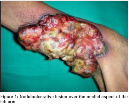
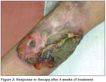
Indian Journal of Dermatology, Venereology and Leprology, Vol. 75, No. 2, March-April, 2009, pp. 199-201 Letter to the Editor Tumor d'emblee responding to methotrexate and prednisolone Aghoram Rajeswari, Thappa DevinderMohan, Kumari Rashmi, Negi VS, Swaminathan RP, Jayanthi S Department of STD, Jawaharlal Institute of Postgraduate Medical Education and Research (JIPMER), Pondicherry - 605 006 Code Number: dv9064 Sir, In 1885, Vidal and Brocq [1] described a rare and unusual variant of mycosis fungoides (MF) called the tumor d′emblee (sudden) type. [1],[2] In this type of MF, the tumors develop suddenly without the usual progression from eczematous to plaque and then tumor stage. Tumors are the initial presentation in approximately 10% of the patients. [1] We herewith report an 83-year-old man presenting with nodular and ulcerative cutaneous T-cell lymphoma (CTCL) with a relatively benign course responding to methotrexate and prednisolone. An 83-year-old man presented with multiple ulcerated lesions on his arms, chest, back and legs of 1 year duration. He noticed multiple nodules that progressively increased in size and ulcerated spontaneously in the center. There was no history suggestive of any preceding skin change or dermatitis. He was initially treated in another hospital as pyoderma gangrenosum with prednisolone (1 mg/kg) to which he had a partial response, but lesions recurred and continued to increase in size and ulcerate. His past medical history was unremarkable. He was a chronic smoker. There was no history of anorexia, weight loss , fever or night sweats. On cutaneous examination, multiple, irregular, hard, mobile, noduloulcerative lesions, largest measuring 13 cm x 10 cm in size, ulcerated in the center and covered with greenish slough were seen over the medial aspect of both arms [Figure - 1], and left forearm, left thenar eminence, and medial aspect of the left flank. The edges of the ulcers were raised and overhanging. The surrounding skin was violaceous and indurated. Two unulcerated nodules were also seen over the back. There was no significant regional lymphadenopathy. Systemic examination was unremarkable and no organomegaly was noted. Histological examination of the skin lesion on the left arm showed a dense infiltrate of mononuclear cells throughout the dermis and subcutis with prominent epidermotropism, with lymphocytes lying singly as well as in groups. Large cell transformation was seen. Immunohistochemistry revealed the tumor cells to be CD3 and leukocyte common antigen positive while being negative for CD30 and CD20. Haemogram, routine blood biochemistry, ultrasound of the abdomen and computerized tomography scan, chest X-ray and urine analysis were normal. Peripheral blood smear, bone marrow aspiration cytology and biopsy examination were negative for atypical cells. Retroviral serology was negative. Pus swab culture and skin biopsy specimen sent for culture from the ulcer grew Pseudomonas aeruginosa sensitive only to amikacin. Based on the clinical and histopathological findings, a diagnosis of tumor d′emblee type of MF (Stage IIb) was made. The patient was treated with a course of antibiotics and daily wet dressing. Subsequently, he was started on single-agent chemotherapy with methotrexate (0.25 mg/kg) weekly along with prednisolone (1 mg/kg) daily. The skin lesions improved slowly and steadily, and after 1 month of treatment, the patient showed regression of more than 70% of the lesions [Figure - 2]. On follow-up after 4 months, only two ulcers remained, which were also healing well without any evidence of dissemination. Primary cutaneous lymphomas represent the second most common extranodal site for non-Hodgkin′s lymphoma. CTCL is a heterogeneous group with diverse clinical manifestations, which represents about 80% of all primary cutaneous lymphomas. [1] The most common CTCL is MF. However, other lymphoproliferative diseases also involve the skin, including Ki-1+ anaplastic large cell lymphoma, peripheral T-cell lymphoma, cutaneous B-cell lymphoma, adult T-cell leukemia/lymphoma, T-cell chronic lymphoid leukemia and cutaneous Hodgkin′s disease. [1],[3] Tumors in MF are usually seen as disease progresses and occur at sites of previous plaque-stage involvement. This progression probably reflects local proliferation and evolution of more aggressive clones. When tumors appear de novo , without preceding plaque or patch disease, they are called tumor d′emblee. [4] It is associated with faster progression and worse prognosis. This was the mode of presentation of our case. Nodular MF that has undergone large cell transformation needs to be closely distinguished from large cell CD30-negative cutaneous lymphoma (peripheral T-cell lymphoma - unclasssified). These tumors also appear suddenly as nodules without pre-existing plaques or patches of MF. On histology, prominent nodular or diffuse infiltrates of medium to large pleomorphic T-cells and immunoblasts are seen but epidermotropism is usually absent. Presence of large CD30-negative pleomorphic T-cells in our case was due to large cell transformation (defined as more than 25-50% of large cells [CD30 positive or negative] within the dermal infiltrate or the development of microscopic dermal nodules of pleomorphic, anaplastic large cells). Transformation has been reported in the range of 8-55% of tumor type of CTCL. [5] Prominent epidermotropism rules out the presence of large cell CD30-negative cutaneous lymphoma (peripheral T-cell lymphoma - unclasssified) in our case. Primary cutaneous (anaplastic) CD30+ large cell lymphomas clinically present with ulcerated nodules mostly on the trunk and can resemble nodular MF but, on histology, no epidermotropism is seen and there is a dense infiltrate of large atypical anaplastic T-cells with CD30+ and frequent mitoses. However, in our case, the cells were CD30 negative and there was no evidence of any systemic involvement thus differentiating this tumor from the anaplastic large T-cell lymphoma. This difference is important to make as the prognosis for a primary cutaneous CD30+ T-cell lymphoma is good as against the large cell transformation of MF. [5] The term ′′tumor d′emblee′′ is now falling into disrepute and these tumors may, in fact, be pleomorphic (small to medium) CD30-negative cutaneous T-cell lymphoma (peripheral T-cell lymphoma), which have undergone large cell transformation. [5] Systemic chemotherapy based on methotrexate with prednisolone was offered to the patient keeping in mind the indolent nature of the disease and the age of the patient, resulting in 70% resolution of lesions. Single-agent chemotherapy with an alkylating agent or methotrexate produces a complete remission rate of 32% and objective remission rate of 63%, with a median duration of remission of 3-22 months. [1] Combination chemotherapy is associated with greater remission rates of up to 80%, but is associated with greater toxicity. [3] References
Copyright 2009 - Indian Journal of Dermatology, Venereology and Leprology The following images related to this document are available:Photo images[dv09064f1.jpg] [dv09064f2.jpg] |
| |||||||||

