 |
| About Bioline | All Journals | Testimonials | Membership | News |
|
||||||
|
||||||
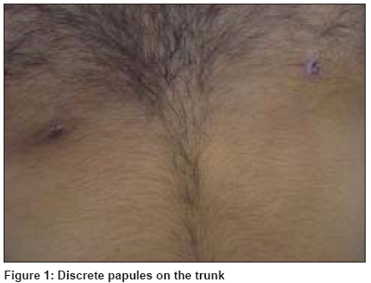
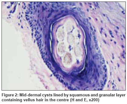
Indian Journal of Dermatology, Venereology and Leprology, Vol. 75, No. 2, March-April, 2009, pp. 217-219 Quiz Asymptomatic papular lesions on the trunk Rao Raghavendra, Balachandran C Department of Dermatology, Kasturba Medical College, Manipal-576 104 Code Number: dv09070 A 24-year-old male presented with asymptomatic skin lesions on the trunk of 6 months duration. There was no history of antecedent trauma or family history of similar illness. Examination revealed discrete tiny skin-colored papules on the anterior chest and abdomen [Figure - 1]. Few lesions showed a comedo-like opening. Biopsy of the lesion showed a middermal cyst lined by flattened epithelium composed of squamous and granular cells surrounding a laminated mass of keratin and multiple vellus hair shafts [Figure - 2]. What is your Diagnosis ? Diagnosis: Eruptive vellus hair cyst (EVHC) Discussion EVHC was first described by Esterly et al . in 1977. [1] It can arise sporadically or in an autosomal-dominant inheritance pattern. In the latter, the disease usually appears at birth or in early infancy, while in the former it begins abruptly without antecedent trauma in the first or second decades of life. [2] There is no sex predilection. The lesions of EHVC are typically small, soft, rounded or dome-shaped, sharply demarcated cystic structures ranging from 1 to 4 mm in diameter. Individual lesions can be flesh-colored, yellow, blue, red or hyperpigmented. A hyperkeratotic crust, central puncta and umbilication have been described in few lesions. [3] Recently, a case of EVHC presenting as facial melanosis resembling naevus of Ota has been described. [4] Generally, they are asymptomatic. Rarely, they may be associated with itching and tenderness. Common sites of involvement are the anterior chest, the abdomen and the extremities. Lesions have also been described on the face, neck, axillae and groin. [5] Histologically, EVHC are characteristically located in the middermis. The epithelium lining the cyst wall is identical to that of the infundibular or sometimes the isthmus portion of the hair follicle and contains two-three layers of squamous epithelium with focal areas of granular layer. The cyst cavity contains variable amount of laminated keratin and multiple transversely cut vellus hairs. Occasionally, the cyst may be surrounded by granulomatous inflammation and the cyst wall may be in continuity with the rudimentary hair follicle or arrector pili muscle. [5] Alternatively, a rapid diagnostic test has been described in which the cyst was incised and the contents of the cyst were expressed. Microscopic examination of the expressed contents in potassium hydroxide preparation showed numerous vellus hairs. [6] The exact cause of EVHC is unknown. Several hypotheses have been postulated. Some consider it to be a hamartoma differentiating toward vellus hair follicle while others believe that cyst arises by blockade of the terminal hair follicle during the stages of development or from faulty development of the infundibulum of the vellus hair follicle. [3] There are reports of EVHC occurring in association with renal failure and with disorders such as anhidrotic ectodermal dysplasia, hidrotic ectodermal dysplasia, pachyonychia congenita and Lowe syndrome. [5] Clinically, steatocytoma multiplex (SM) mimics EVHC; however, there are subtle histopathological differences. SM consists of an empty cyst covered by a thin wall of stratified squamous epithelium. The main feature is the presence of sebaceous elements within or adjacent to the cyst wall. In contrast, EVHC consists of a small cyst formed by several layers of squamous cells and a cavity that contain vellus hairs. [7] Jerosutus et al . believe that both conditions are spectrum of the same disease that represents developmental anomaly from different portions of the hair follicle. [8] Gonzalez et al . described a hybrid cyst showing histopathological characteristics of both SM and EVHC. [9] However, Tamkova et al. revealed, through staining with K 10 and K 17, that EVHC and SM were in fact two distinct entities. [10] Other differential diagnosis includes milia, trichilemmal cyst, dermoid cyst, perforating collagenosis, acne comedones and epidermal inclusion cysts. Patients generally seek medical attention for cosmetic reasons. Treatment of EVHC is often challenging, with disappointing results. Topical therapies with 10% urea, 12% lactic acid, retinoic acid and 0.1% tazarotene have yielded favorable results in few patients. Simple and destructive surgical techniques, which enable rapid treatment of multiple lesions, have been described; however, their utility is limited due to inconsistent results. [11] Approximately 25% of the EVHC cases resolve spontaneously by extruding their content. References
Copyright 2009 - Indian Journal of Dermatology, Venereology and Leprology The following images related to this document are available:Photo images[dv09070f1.jpg] [dv09070f2.jpg] |
| |||||||||
