 |
| About Bioline | All Journals | Testimonials | Membership | News |
|
||||||
|
||||||
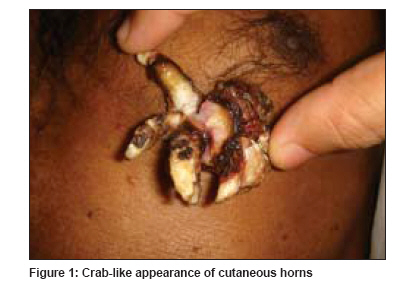
Indian Journal of Dermatology, Venereology and Leprology, Vol. 75, No. 3, May-June, 2009, pp. 300-301 Images in Clinical Practice Crab-like appearance of cutaneous horns Nath AmiyaKumar, Thappa Devinder Mohan Department of Dermatology and STD, Jawaharlal Institute of Postgraduate Medical Education and Research (JIPMER), Pondicherry - 605 006 Code Number: dv09088 PMID: 19439887 A 65-year-old man presented with an asymptomatic pedunculated growth on the chest since two years. Initially nodular, verrucous growth slowly enlarged, and developed into multiple horns-like projections. Cutaneous examination revealed a pedunculated growth measuring 3 x 3 cm on the chest, with five well-developed horns arising from it, appearing like a crab sitting on the chest [Figure - 1]. The growth had a pedicle of diameter 1 cm, withan indurated, tender base of size 1.5 x 1 cm. No regional lymphadenopathy was noted. The result of systemic examination was within normal limits. The entire lesion including the indurated base was excised with an adequate margin. Histopathological examination of the excised mass revealed well-differentiated squamous cell carcinoma in the bases of the horns, showing superficial invasion in broad tongues. Deep margin and lateral margins of the resection were free of tumor. Cutaneous horn (cornu cutaneum) - named so because of its resemblance to animal horn - is a conical, hyperkeratotic, protuberant mass composed of compact keratin caused by increased adhesiveness of the keratinized material. However, the animal horns are composed of superficial hyperkeratotic epidermis and dermis with centrally positioned bone. No such well-formed bone is observed in human horns. Cutaneous horns present as a hard, yellowish brown protrusion, often curved and have circumferential ridges, which are surrounded by either normal-appearing epidermis or an acanthotic collarette. The height of a cutaneous horn is at least half of its diameter at the base. They are usually single, but multiple horns may occur. They most frequently occur in sun-exposed parts and are typically found on the face and scalp, but may also occur on the hands, penis, eyelids, nose, chest, neck and shoulder. Cutaneous horn has been described overlying a wide variety of benign, premalignant and malignant conditions such as seborrheic keratoses, nevus, wart, molluscum contagiosum, rhinosporidiosis, psoriasis, lichen planus, porokeratosis, actinic keratosis, histiocytoma, follicular infundibulum tumor, keratoacanthoma, basal cell carcinoma and squamous cell carcinoma. The horn may vary from a few millimeters to several centimeters (including giant horns) in size, and have flat, nodular or crateriform base. Inflammation may occur due to recurrent injury. However, inflammation, induration and tenderness at the base of the lesion and lesions of larger size favor malignancy. Histologically, a horn consists of greatly thickened stratum corneum, with scattered areas of parakeratosis. The granular layer may be deficient or absent. The horn at the base usually displays the features characteristic of the pathologic process responsible for the development of the horn. A majority of cutaneous horns are associated with benign pathology. Malignancy may be present in 16%-20% of cases, with squamous cell carcinoma being the most common type. However, 33% of penile cutaneous horns may be associated with squamous cell carcinoma. In Mencia-Gutierrez et al′s retrospective analysis of 48 cutaneous horns, 77.1% of the horns were associated with a benign pathology, 14.6% with a premalignant and 8.3% with a malignant lesion. Seborrheic keratosis was the most common among the benign lesions, actinic keratosis among the premalignant lesions and basal cell carcinoma and squamous cell carcinoma among the malignant lesions. Because they are suggestive of underlying anaplasia, biopsy examination is warranted. The crab-like cutaneous horn in our case was unique in its appearance. Copyright 2009 - Indian Journal of Dermatology, Venereology and Leprology The following images related to this document are available:Photo images[dv09088f1.jpg] |
| |||||||||
