 |
| About Bioline | All Journals | Testimonials | Membership | News |
|
||||||
|
||||||
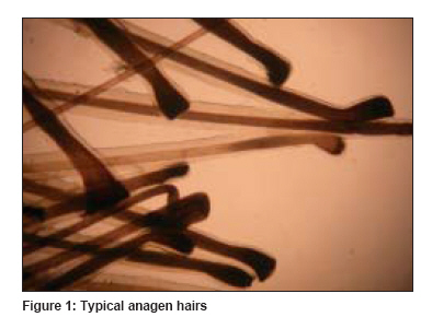
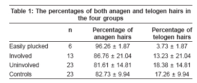
Indian Journal of Dermatology, Venereology and Leprology, Vol. 75, No. 3, May-June, 2009, pp. 303-304 Letter to the Editor Trichogram findings in pemphigus patients Koslu Adem, Topal IlterisOguz, Ekmekci TugbaRezan Department of Dermatology, Sisli Etfal Research and Training Hospital, Istanbul Code Number: dv09090 PMID: 19439889 Sir, Pemphigus is an autoimmune blistering disease in which an antibody interacts with desmosomal proteins, leading to acantholysis of epidermal cells. One of the preferential targets of pemphigus autoantibodies is hair follicle, as the desmosomal proteins are overexpressed in follicular epithelium. Moreover, direct immunofluorescence of the outer root sheath of plucked hair is a reliable and specific method for diagnosing pemphigus. [1],[2] The changes in the hair follicle in pemphigus have been shown in several immunofluorescence and electron microscopic studies. However, to date, only one study has been performed by trichogram. [3] Recently, a different trichogram finding has been reported in pemphigus. [4] Our aim was to discover whether there is a different trichogram finding in pemphigus and, if so, how frequent its incidence is. A total of 23 pemphigus patients (15 men and 8 women, mean age: 51.17 ± 15.37 years) who were newly diagnosed by histopathology and direct immunfluorescence and did not receive any treatment and 23 controls (15 men 8 women, mean age: 52.52 ± 15.20 years) were included in the study. Scalp involvement was accepted as positive only when vesicles, pustules, erosion, crusted erythematous plaques, plaques with pustules were present on the scalp. When scalp involvement was found, hair was plucked from both the involved area and the occipital scalp. If not, hair was plucked only from the occiput. The percentages of both anagen and telogen hairs were estimated. Data analysis was performed using the SPSS 11.0 (SPSS Inc., USA). Statistical analysis was accomplished between the groups, using Students t test and Mann-Whitney U test. Values were reported as mean ± SD; statistical significance was attributed to two-tailed P < 0.05. Pemphigus vulgaris and pemphigus vegetans were diagnosed in 21 (91.3%) and 2 patients (8.69%), respectively. The mean duration of the disease was five months. The disease was severe in 2 patients, moderate in 16 and slight in 5. Scalp involvement was observed in 13 patients (56.52%) (involved group). No alopecia was noted in the involved areas. Of the13 patients, in 6 (46.15%), the hairs were plucked very easily without any resistance and almost all of them were typical anagen hairs with intact root sheats [Figure - 1] (easily plucked group). The percentages of both anagen and telogen hairs of four groups (easily plucked vs. involved vs. univolved vs. controls) are shown in [Table - 1]. When the percentages of anagen hairs were compared among these four groups, the percentage of anagen hairs in easily plucked group was statistically higher than that in the others (respectively, P = 0.31, P = 0.30, P = 0.44) and that of telogen hairs was also statistically lower (respectively, P = 0.31, P = 0.30, P = 0.44). Furthermore, while in easily plucked group there were no dystrophic or dysplastic anagen hairs, in the remaining groups they were present. Delmonte et al ., [4] described normal anagen effluvium in three pemphigus patients. They reported that all the hairs were easily plucked and resembled normal anagen hairs with root sheats similar to those in our easily plucked group. They stated that the normal anagen effluvium suggested a subclinical involvement of the hair follicle and that it could be considered as a Nikolsky′s sign of the scalp. Afterward, one additional pemphigus case with normal anagen effluvium was reported. [2] We also think that normal anagen effluvium is a Nikolsky′s sign of the scalp. It would be better if the clinicians pay attention to this important clinical finding that has been overlooked so far, especially in pemphigus cases either confined to or initially starting from the scalp.The hairs were easily plucked because in pemphigus, intraepidermal acantholytic blister formation with acantholysis often extends from the epidermis to down the outer root sheats of the hair follicles. [3] In the first trichogram study composed of 10 pemphigus patients, the authors described an increase in the number of telogen hairs in pemphigus vegetans and pemphigus seborrheicus and that normal trichogram findings in pemphigus vulgaris and foliaceus were seen. They did not mention about normal anagen effluvium or easily plucked hair. [4] We regard that if scalp involvement is not observed in pemphigus, trichogram will neither be a beneficial nor a helpful method for the clinician. The percentage of telogen hairs was low in our easily plucked group. Desmoglein 3 seemed to be of major importance for anchoring the telogen club hair to the outer root sheath. [5] This hypothesis can explain the low telogen percentages as a result of early telogen hair loss. As the plucking technique may produce dystrophic or dysplastic anagen hairs, [6] we did not calculate these hairs seperately in anagen hairs. However, while we observed only typical anagen hairs in easily plucked group, we saw dystrophic and dysplastic anagen hairs in the others. The reason for this may be explained that the hairs come out easily without any break at different levels in the hair follicle. References
Copyright 2009 - Indian Journal of Dermatology, Venereology and Leprology The following images related to this document are available:Photo images[dv09090t1.jpg] [dv09090f1.jpg] |
| |||||||||

