 |
| About Bioline | All Journals | Testimonials | Membership | News |
|
||||||
|
||||||
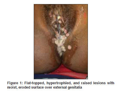
Indian Journal of Dermatology, Venereology and Leprology, Vol. 75, No. 4, July-August, 2009, pp. 401-402 Images in Clinical Practice Moist flat-topped raised genital lesions in late pregnancy Nath AmiyaKumar, Rajalakshmi R, Thappa DevinderMohan Department of Dermatology and STD, Jawaharlal Institute of Postgraduate Medical Education and Research (JIPMER), Pondicherry - 605 006 Code Number: dv09153 PMID: 19584469 A 26-year-old primigravida in seventh month of pregnancy presented with multiple, asymptomatic, raised lesions over the vulva, perineum and peri-anal regions of 4 weeks duration. Her husband had a few small raised lesions near the anus. Neither of them had fever, headache, myalgia, arthralgia or skin lesions elsewhere. There was no past history of genital ulcers in either spouse. There was no history of sexual promiscuity in the patient, but her husband had multiple, unprotected, pre-marital homosexual and heterosexual contacts seven years back. On examination of the external genitalia of the patient, there were multiple, discrete-to-coalescing, flat-topped, hypertrophied, raised lesions with moist, eroded surface over the labia majora, vaginal introitus, fourchette, perineum, groin and peri-anal region [Figure - 1]. Firm, non-tender, mobile, bilateral inguinal lymphadenopathy was present. There was no epitrochlear lymphadenopathy or lymphadenopathy at other sites. Other mucosal, cutaneous and systemic examinations were within normal limits. Examination of the husband revealed two small condyloma acuminata lesions around the anal margin. No other abnormality was noted in the husband. Serological investigations in both of them revealed a reactive VDRL (Venereal Disease Research Laboratory) test in 1:32 dilution, and a positive Treponema Pallidum Haem Agglutination (TPHA). ELISA for HIV-1 and 2 was negative in both. Histopathology of one excised lesion from the patient showed hyperplastic epidermis, parakeratosis, spongiosis, intra-epidermal neutrophils collections, papillary dermal edema, peri-vascular and peri-adnexal lymphohitiocytic and plasma cell infiltrates, and a few areas of epithelioid cell granulomas in the dermis. Warthin-Starry stain demonstrated numerous treponemes in the biopsy specimen. A final diagnosis of condyloma lata was made, and the patient and her husband were treated with single intra-muscular injection of benzathine penicillin 2.4 million units. The condyloma lata lesions started reducing in size two days after the injection and completely disappeared after six days. The husband was also treated for peri-anal condyloma acuminata with cryotherapy. Condylomas are papular syphilids, essentially, vegetative hypertrophy of the epidermis characteristically seen in secondary syphilis. They are confluent, hypertrophic, pale-colored broad and flattened papules occurring in warm moist areas. They begin as raised, slightly mammillated or smooth papules with moist eroded surfaces and are more common in women rather than men due to moisture and friction in female genitalia favoring their development. In women, the papules occur more commonly around the anus, the vulva and the cervix. In some patients, the papules may show a predilection for certain sites such as corners of the mouth, angles of the nose, palms and soles and body folds such as beneath the breasts and axillae. Rarely, they may involve the face, neck, umbilicus, toe webs and eyelids. The mucous lesions have an evanescent character. (i.e., they may be here today and gone tomorrow). They may rarely become pedunculated. Linear condylomas may develop in flexures. Neighboring lesions may become confluent forming soft spongy masses with a cauliflower structure obscured by the erosion of the surface and by a thin grayish pellicle of exudates. Condyloma lata are highly infectious and teeming with spirochaetes. During the later stages of pregnancy, hypertrophic, coalesced, sodden-surfaced papules may be very pronounced. Copyright 2009 - Indian Journal of Dermatology, Venereology and Leprology The following images related to this document are available:Photo images[dv09127f1.jpg] |
| |||||||||
