 |
| About Bioline | All Journals | Testimonials | Membership | News |
|
||||||
|
||||||
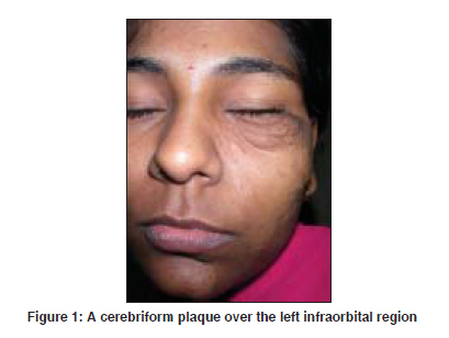
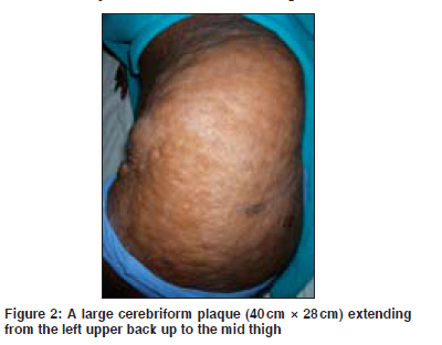
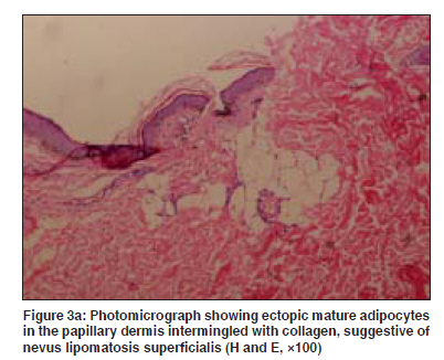
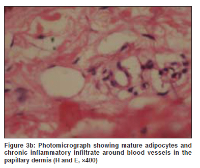
Indian Journal of Dermatology, Venereology and Leprology, Vol. 75, No. 4, July-August, 2009, pp. 407-408 Letter to the Editor Giant nevus lipomatosus cutaneous superficialis Khandpur Sujay, Nagpal SmitaA, Chandra Subhash, Sharma VinodK, Kaushal Seema, Safaya Rajni Department of Dermatology and Venereology, All India Institute of Medical Sciences, New Delhi Code Number: dv09157 PMID: 19584473 Sir, Nevus lipomatosus cutaneous superficialis (NLCS), first described by Hoffman and Zurhelle in 1921, is a rare dermatosis characterised by multiple, variable-sized, flesh-colored to yellowish sessile plaques with cerebriform surface or small solitary nodules due to nevoid fatty growth within the papillary and reticular dermis. [1] We report two plaques of NLCS in an 18-year-old woman, one over the left face and the other extending from the upper back to the thigh (giant NLCS). This case is being reported for the rarity of the disorder and the large size of the lesion. As far as ascertained, the second plaque is the largest lesion being reported in the literature. An 18-year-old woman presented with two asymptomatic, skin-colored cerebriform plaques over the left side of the face and neck and the left upper back extending to the mid thigh since 10 years of age. The face lesion was preceded by trauma. She presented to us for cosmetic reasons. There were no systemic complaints. Dermatological examination revealed two large plaques, the first 13.5cm x 4cm in size, composed of multiple skin-colored, soft, coalescent papules over the left side of the face and neck with a skin laxity below the left eyelid [Figure - 1] and the second, a large cerebriform plaque 40cm x 28cm in size, extending from the left upper back and flank to the antero-lateral aspect of the mid thigh [Figure - 2]. The overlying skin was nonhairy and normally pigmented. Skin biopsies from both the thigh and the neck showed collection of mature adipocytes intermingled with collagen bundles in the papillary and upper reticular dermis. The adipocytes along with chronic inflammatory infiltrate were also present around the blood vessels. The histological features were compatible with nevus lipomatosus [Figure - 3]a and b. On magnetic resonance imaging, abnormal areas of fat deposition on the left side of the face, left flank and thigh in the subcutaneous plane with no infiltration of the internal organs and muscles were seen. The patient was referred to the plastic surgery department for surgical excision. NLCS is a rare disorder characterized by nevoid accumulation of mature adipocytes in the dermis. It often presents at birth but can appear within the first two decades of life, as in our case. [2] There is no familial or sex predilection. Two clinical types are recognized. The classical type, reported by Hoffmann and Zurhelle, consists of multiple flesh-colored or yellowish sessile lesions with a tendency to coalesce into plaques with a smooth or corrugated surface and zonal distribution and following natural cleavage lines of the skin. They have a predilection for the pelvic girdle and sacral and lumbar regions. The second type manifests as small solitary nodules mimicking skin tags and occurring over the arms, knees, axillae, ears and scalp. They appear at a later stage of life. Occurrence over atypical sites like the nose, calf and clitoris has also been reported. In our patient, both the plaques were of the classical type, the second one being a giant NLCS, 40cm x 28cm in size. The largest size reported so far has been a 20cm x 30cm-sized bilateral cerebriform plaque extending from the lower abdomen to the left leg in a 36-year-old Brazilian woman. [3] NLCS has been reported in association with cafι au- lait macules, leucodermic spots, hypertrichosis over the nevus, comedo-like lesions and angiokeratoma of Fordyce. [4] The pathogenesis of NLCS is unknown and several theories have been put forth. Proposed pathogenesis include adipose metaplasia during the course of degenerative changes in dermal connective tissue, as stated by Hoffman and Zurhelle and supported by Nikolowsky, and developmental displacement (heterotopia) of adipocytes or development of mature adipocytes by mesenchymal perivascular cells present initially as primitive lipoblasts that subsequently transform into mature adipocytes. [2] On histopathology, the distinguishing feature is presence of ectopic mature adipocytes in the dermis intermingled with collagen bundles and perivascular infiltration of dermis and subcutis with chronic inflammatory cells. [2] This condition may be confused with plexiform neurofibroma, connective tissue nevus, vascular malformation, lipomatosis or lipoblastomatosis. The treatment of choice is surgical excision. [5] Patients unwilling for surgery may undergo cryotherapy, which yields partial though satisfactory results. [4] References
Copyright 2009 - Indian Journal of Dermatology, Venereology and Leprology The following images related to this document are available:Photo images[dv09131f2.jpg] [dv09131f3a.jpg] [dv09131f1.jpg] [dv09131f3b.jpg] |
| |||||||||
