 |
| About Bioline | All Journals | Testimonials | Membership | News |
|
||||||
|
||||||
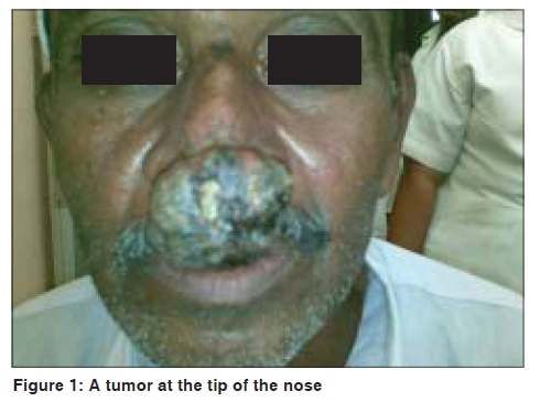
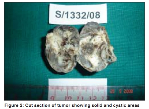
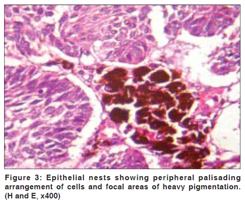
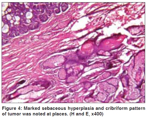
Indian Journal of Dermatology, Venereology and Leprology, Vol. 75, No. 5, September-October, 2009, pp. 506-508 Case Report Pigmented trichoblastoma with sebaceous hyperplasia Girish Kamat, Balasab Yelikar, Savita Shettar, Mahesh H. Karigoudar Department of Pathology, Shri B. M. Patil Medical College and Research Centre, Bijapur, India Code Number: dv09159 PMID: 19736433 DOI: 10.4103/0378-6323.55397 Abstract Trichoblastoma is a rare benign trichogenic tumour with epithelial and mesenchymal components recapitulating the germinal hair bulb and associated mesenchyme. A 50- year- old male patient presented with slowly enlarging circumscribed solid nodule measuring 4x3x4.5 cm over the tip of the nose. Microscopy of tumour revealed nodular tumour spanning the entire dermis with collection of mesenchymal cells resembling follicular papilla. Areas of pigmentation and sebaceous hyperplasia were noted. There is a need for differentiation of this tumor which is benign, from other pigmented tumors having basaloid arrangement of cells such as basal cell carcinoma.Keywords: Trichogenic tumors, Pigmented trichoblastoma, Pigmented epithelial tumors Introduction Trichoblastoma presents clinically as slowly growing, solitary, well circumscribed nodule, predominantly in head and neck area. Most commonly it is seen in age group of 5 th to 7 th decade. [1] Trichoblastoma is the most common neoplasm developing in nevus sebaceous of Jadassohn. [2] Rare case of trichoblastoma with sebeceoma has been reported. [3] A rare case of trichoblastoma with apocrine and sebaceous differentiation also has been reported. [4] There are very few cases of pigmented trichoblastoma reported in the literature. [5] To the best of our knowledge, pigmented trichoblastoma with sebaceous hyperplasia has not been described in the literature. Case Report A 50-year-old male patient, presented to Ear, Nose and Throat department with a gradually enlarging mass over the tip of nose for last 6 years. The nodule was well circumscribed and ovoid. The surface was smooth and black to brown in colour [Figure - 1]. It was firm in consistency and was tender to palpate. A clinical differential diagnosis of benign adnexal tumour, keratinous cyst, and pigmented basal cell carcinoma was made and the patient was referred for fine needle aspiration cytology. Fine needle aspiration cytology revealed clusters of basaloid cells with elongated euchromatic nuclei and scant cytoplasm. A provisional diagnosis of benign adnexal tumor was made and simple excision of mass was carried out. On gross examination, tumour was of 4x3x4.5cm in size, partially capsulated, and was greyish brown in colour. On cut section, mass showed solid and cystic areas. Solid areas were greyish brown in color. Cystic areas showed hemorrhage and necrosis [Figure - 2]. On microscopic examination, well circumscribed nodular tumor was noted spanning the entire dermis. At scanning magnification, multiple cerebriform lobules of various sizes and patterns were noted. The lobules were not connected with the overlying epidermis and were separated by collagen. Peripheral palisading was conspicuous but there was no stromal condensation around the tumor lobules and cleft artefact was not a prominent feature. At places cribriform pattern was noted. Also larger lobules showed keratin cyst formation, although much less frequently than seen in trichoepithelioma. Atypia and mitotic figures were found to some extent. There was no cell necrosis and local infiltration, as is normally observed in a malignancy. In addition to these features, there were areas of heavy pigmentation, which contained abundant intralesional dendritic melanocytes [Figure - 3]. Adjacent to the tumor tissue, marked sebaceous hyperplasia was noted [Figure - 4]. Based on the overall histopathology features, a diagnosis of pigmented trichoblastoma with sebaceous hyperplasia was made. Discussion Trichoblastoma is a benign neoplasm with primitive hair follicle formation. The tumor is characterized by nests and cords of epithelial cells in an organised relationship with stroma. This condition was defined as tumor of hair germ by Headington in 1970. He divided this group of tumors into trichoblastoma, and trichogenic myxoma based on the nature of the stroma and the degree of "induction", specifically, a collection of mesenchymal cells resembling the follicular papilla in a close approximation to the epithelial cells similar to those of the hair germ or the inner or outer root sheath. [6] Usually most trichoblastomas develop along only one line of differentiation (i.e. toward primitive hair follicle). Rare cases of trichoblastoma show multiple paths of differentiation toward more than one type of adnexal structure. [3],[4] Yu et al . have reported a case of trichoblastoma which showed apocrine and sebaceous differentiation. [4] From this point of view, the presence of sebeceous differentiation in trichoblastoma as in our case might be explained by the phenomenon of multidirectional differentiation involving the pleuripotent cells with an adnexal structure. These findings are consistent with the theory of a common embryological origin for the three adnexal components as a folliculo-sebeceous-apocrine unit and eccrine gland from embryonal stratum germinatum. [7] The close differential diagnosis for pigmented trichoblastoma is pigmented basal cell carcinoma. But basal cell carcinoma arises from epidermis and later invades into the dermis. Differentiation toward the hair germ is not found, and the pigmentation is restricted to the upper part of dermis. [5] In 1999, Kaddu et al . have reported 2 cases of trichoblastoma and sebeceoma in nevus sebeceous. [3] Aloi et al . also have reported a case of pigmented trichoblastoma. [5] To conclude, we have described an unusual variety of trichogenic tumor with pigmentation and sebaceous differentiation. To the best of our knowledge such a tumor has not been described before in the literature. References
Copyright 2009 - Indian Journal of Dermatology, Venereology and Leprology The following images related to this document are available:Photo images[dv09159f2.jpg] [dv09159f4.jpg] [dv09159f1.jpg] [dv09159f3.jpg] |
| |||||||||
