 |
| About Bioline | All Journals | Testimonials | Membership | News |
|
||||||
|
||||||
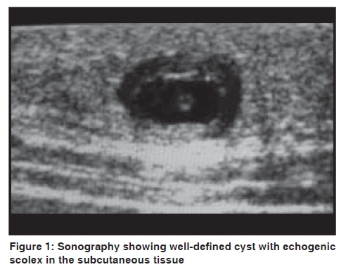
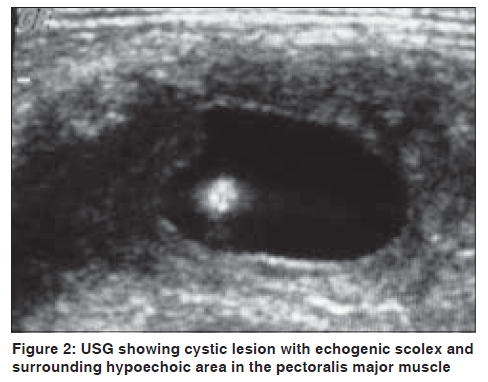
Indian Journal of Dermatology, Venereology and Leprology, Vol. 75, No. 5, September-October, 2009, pp. 515-516 Letter to the Editor Subcutaneous and intramuscular cysticercosis: High-resolution sonography Amit Mittal, Sanjeev Gupta1, Sunita Gupta2, Vinod Mehta Departments of Radiodiagnosis, 1Dermatology and 2Medicine, MM Institute of Medical Sciences and Research, Mullana, Ambala, India Code Number: dv09166 PMID: 19736438 DOI: 10.4103/0378-6323.55404 Sir, We read the article 'Subcutaneous nodules preceding convulsions due to neural cysticercosis' by Singrodia et al . [1] They had rightly pointed out that the prerequisite for successful treatment of cysticercosis with anthelminthics is early diagnosis; however, they did excisional biopsy for the diagnosis. Here, we want to draw the kind attention to a very important non-invasive diagnostic technique for subcutaneous and intramuscular cysticercosis i.e. high-resolution sonography. High-resolution sonography is a diagnostic investigation for subcutaneous and intramuscular cysticercosis. [2],[3] We are discussing here two patients with subcutaneous and intramuscular cysticercosis. First patient was a 11-year-old male who presented with an asymptomatic swelling on the left side of the chest of one-month duration. Clinically, it was diagnosed as lipoma or neurofibroma. On high-resolution sonography (done on Logiq 500 PRO machine, GE Medical Systems, with a linear probe at 9.6 MHz frequency), there was a well-defined cystic lesion of size 12 × 11 mm with echogenic nidus in the subcutaneous tissue in the area of swelling, suggestive of cysticercosis [Figure - 1]. The second patient was a 35-year-old male presenting with a painful swelling of the anterior chest wall for last 8-10 days on the right side. On examination, the swelling (approximately 3 × 3 cm) was tender and hard and skin overlying the swelling was inflamed. There was no history of fever or trauma. Clinically it was diagnosed as an abscess. On high-resolution sonography there was a cyst of size 14 × 12 mm with small 3 mm echogenic scolex in it with surrounding hypoechoic area of size 25 × 22 mm in the left pectoralis muscle suggestive of cysticercosis [Figure - 2]. In both these cases, no further invasive or immunological investigations were done for the diagnosis. They were treated with albendazole and corticosteroids and recovered completely. Therefore, high-resolution sonography plays an important role in the diagnosis of muscular and subcutaneous cysticercosis. On ultrasound, cysticercosis can appear as the cyst with an eccentric echogenic scolex and with an inflammatory mass around it, because of the death of the larva. It can appear as an irregular cyst without echogenic scolex and with minimal fluid on one side, indicating the leakage of fluid. The echogenic scolex is not seen within the cyst because it might escape outside the cyst, or because of the partial collapse of the cyst. The third appearance is a large irregular collection of exudative fluid within the muscle, with the typical cysticercus cyst containing the scolex situated eccentrically within the collection. This may be due to chronic intermittent leakage of fluid from the cyst, leading to florid inflammatory exudates. This appearance is similar to an intramuscular abscess, but the visualization of the cysticercus cyst within it clinches the diagnosis. In all three of these types, the salient diagnostic feature is that of the cysticercus itself, which appears as an oval or round well-defined cystic lesion with an eccentric echogenic scolex in it. The fourth ultrasound appearance is that of calcified cysticercosis. [2],[3] These appearances on high-resolution sonography are pathognomonic of cysticercosis, and a definitive diagnosis can be made with greater confidence and patient can be managed conservatively. Therefore, with the help of non-invasive high-resolution sonography such subcutaneous and intramuscular cysticerci can be accurately diagnosed without need of invasive biopsy or fine needle aspiration cytology (FNAC). Further, the diagnosis of sonography findings can be confirmed with therapeutic response as was also done in our cases. [4] By this report, we want to make the dermatologists and surgeons aware of high-resolution sonography as an important non-invasive tool for the proper diagnosis and management of subcutaneous/intramuscular swellings. References
Copyright 2009 - Indian Journal of Dermatology, Venereology and Leprology The following images related to this document are available:Photo images[dv09166f1.jpg] [dv09166f2.jpg] |
| |||||||||

