 |
| About Bioline | All Journals | Testimonials | Membership | News |
|
||||||
|
||||||
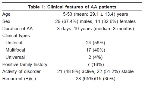
Indian Journal of Dermatology, Venereology and Leprology, Vol. 75, No. 5, September-October, 2009, pp. 552 Net letter Serum vitamin B12, folate, ferritin, and iron levels in Turkish patients with alopecia areata Müzeyyen Gönül, Seray Külcü Çakmak, Seçil Soylu, Arzu Kõlõç, Ülker Gül Ankara Numune Education and Research Hospital, 2nd Dermatology Clinic, Ankara, Turkey Code Number: dv09191 PMID: 19736464 DOI: 10.4103/0378-6323.55430 Sir, Alopecia areata (AA) is a nonscarring inflammatory hair loss on the scalp and/or body with unclear etiopathogenesis. There are only a few reports investigating serum iron and ferritin levels in AA patients. [1],[2],[3] The levels of vitamin B12, folate in AA have not been investigated in AA patients. We aimed to investigate vitamin B12, folate, iron, and ferritin levels in AA patients and their potential role in the etiology of AA. A total of 43 AA patients and 36 healthy sex and age-matched individuals were retrospectively included in the study within eight-month period, in 2008. Age, sex, duration, activity, and family history of AA were obtained from the previous records. Exclusion criteria were other autoimmune disorders, pregnancy, drug usage, and other associated diseases that could alter levels of vitamin B12 ferritin, iron and folate. The patients were divided into two groups the stable and active according to the progression of the lesions in the last month. Also, the disease was divided into three groups as unifocal, multifocal, and universal according to the clinical appearance. Serum hemoglobin, iron (autoanalyser, Olympus AU 800 Japan), vitamin B12, folate, and ferritin (chemiluminisence immunoassay/microparticle, Abbot, Germany) levels of the subjects were obtained from the medical records. Lower hemoglobin level than normal for the sex was accepted as anemia. The levels of vitamin B12, folate, ferritin, and iron in AA patients were compared with those of controls and the parameters of AA were also compared with the sex of patients, duration, activity, and family history of AA. Clinical subtypes of AA were compared with controls and with each other. Statistical analysis was carried out using Mann-Whitney U test, Spearmann correlation analysis, and Kruskal-Wallis test. Out of the total AA patients, 29 (67.4%) were male and 14 (32.6%) were female. The clinical features of AA patients are shown in [Table - 1]. Low levels of hemoglobin, vitamin B12, ferritin, folic acid, and iron were detected in 3, 4, 4, 2, and 8 AA patients, respectively. No significant difference was found between the patient and the control groups in hemoglobin, vitamin B12, ferritin, folate, and iron levels. Folate, iron, and vitamin B12 levels did not show significant variation in relation to family history, duration, and activity of disorder. Ferritin and hemoglobin levels were significantly lower in females than that in males in the patient group ( P < 0.001, P < 0.004, respectively). There was no significant difference when the clinical subtypes of AA were compared with controls and with each other. Many theories have been suggested in the etiopathogenesis of AA including infectious, neural, genetic, and organ specific autoimmune hypotheses. There is increasing evidence that AA is a tissue-specific autoimmune disease. AA has been associated with various autoimmune disorders. [1],[2],[3] To our knowledge, our study is the first study investigating vitamin B12 and folate levels in the patients with AA. In our study, vitamin B12 and folate levels were not different in AA patients from the control group. Another probable mechanism is oxidative stress characterized by an increase in free radical production exceeding the intracellular antioxidant defense. Akar et al. suggested that lipid peroxidation and antioxidant enzymes might be related to the pathogenesis of AA. [4] Iron, the essential element for many important cellular functions, is involved in antioxidative system and large amount of iron is sequestered by the ferritin. The expression of ferritin is regulated by the levels of iron, cytokines, hormones, and oxidative stress. Ferritin has been reported to exhibit different immunological activities such as suppression of antibody production by lymphocytes and suppression of delayed type hypersensitivity. The ferritin levels are increased in inflammation, infections, and malignancies. [5] Recently, ferritin has been accepted as a novel marker for autoimmunity and elevated levels of ferritin in autoimmune disorders have been reported. Ferritin and iron levels have been evaluated in a few studies in AA patients. While White et al. concluded that female AA patients have an increased incidence of iron deficiency in comparison with the general population, Boffa et al . and Esfandiarpour et al. suggested that the prevalence of iron deficiency is not significantly increased in patients with AA. [1],[2],[3] Our study has supported the results of Boffa et al. and Esfandiarpour et al . In this study, serum ferritin, iron, vitamin B12, and folate levels in AA patients were not different from than that in the control group. The levels of serum ferritin, iron, vitamin B12, and folate did not vary according to duration of disorders or the activity of AA. Absence of significant differences may be due to the small sample size. We think that iron, ferritin, vitamin B12, and folate do not play a role in the etiology and activity of AA. References
Copyright 2009 - Indian Journal of Dermatology, Venereology and Leprology The following images related to this document are available:Photo images[dv09191t1.jpg] |
| |||||||||
