 |
| About Bioline | All Journals | Testimonials | Membership | News |
|
||||||
|
||||||
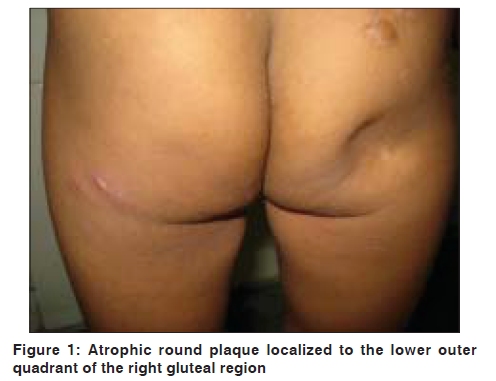
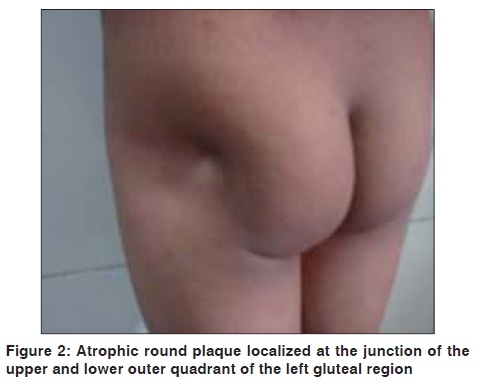
Indian Journal of Dermatology, Venereology and Leprology, Vol. 75, No. 5, September-October, 2009, pp. 552 Net letter Localized lipoatrophy after intramuscular amikacin Vishal Kumar, Manish Kumar, Chander Grover1 Departments of Pediatrics and 1Dermatology, Venereology, and Leprology, Maulana Azad Medical College and Associated, Chacha Nehru Bal Chikitsalaya, Delhi, India Code Number: dv09192 PMID: 19736463 DOI: 10.4103/0378-6323.55431 Sir, Localized acquired lipoatrophies constitute a poorly defined group of diseases clinically characterized by focal loss of fat. Many cases may be idiopathic, characterized by loss of adipose tissue without antecedent clinical or histologic inflammation. [1] Lipoatrophy may also occur as a consequence of inflammatory diseases and repeated trauma such as drug injections. Drugs that have been implicated in the pathogenesis of lipoatrophy are insulin, penicillin, and corticosteroids. [1],[2],[3] We report two cases of acquired localized lipoatrophy after multiple intramuscular injections of amikacin, which have not been reported in the literature yet. A detailed history and examination did not reveal any antecedent cause of lipoatrophy in both cases. Case 1: A 3-year-old male child presented with nonprogressive localized depression in the right gluteal region, which was noticed one month ago. He had acute diarrhea five months back (four months prior to the appearance of the gluteal depression) for which he received intramuscular amikacin injections over five days, with most of the injections administered in the right gluteal region. No other parenteral medication was given. There was no history of pain, redness, or swelling at the site during or after the course of injections. He never received any other intragluteal injection. On examination, a depressed circular plaque, 3.7 cm in diameter with some hypopigmentation of the overlying skin, was seen in the lower outer quadrant of the right gluteal region [Figure - 1]. There was no sign of inflammation. A scar mark suggestive of a healed folliculitis was also present in the same gluteal region, in the upper outer quadrant. No systemic side effects of amikacin were observed. Complete blood counts, erythrocyte sedimentation rate, renal function tests, and urine routine examination were within normal limits. Antinuclear antibody (ANA), anti-dsDNA, anti-Sm, and anti-U1-RNP antibodies were negative by qualitative immunoassay. Histopathology showed a decrease in the number and size of adipocytes, with few perivascular mononuclear cells. No treatment was prescribed and on follow up after three months, an examination revealed signs of resolution. Case 2: A 6-year-old boy presented with nonprogressive localized depression in the left gluteal region, noticed two months ago. He had febrile urinary tract infection four months back (two months prior to the gluteal depression), for which he received multiple intramuscular amikacin injections over seven days, with most of the injections administered in the left gluteal region. No other parenteral medication was given. There was no history of pain, redness or swelling at the site after the course of injections. He never received any other intragluteal injections. On examination, a hypopigmented, depressed, circular plaque, 4.3 cm in diameter, was seen at the junction of the upper and lower outer quadrant of the left gluteal region [Figure - 2]. Similar to the first case, no signs of inflammation at the injection site and no systemic side effects of amikacin were observed. All blood and urine investigations, as performed in the first case, were within normal limits. No treatment was prescribed and on follow up after two months, an examination revealed signs of resolution. The terms lipoatrophy and lipodystrophy are often used interchangeably and without precise definition. Lipodystrophy is best defined as an abnormality of fat that is usually associated with atrophy, or infrequently hypertrophy of the adipose tissue. [4] Lipoatrophy refers specifically to the loss of fat. Lipoatrophy can be localized, partial, or generalized. Localized lipoatrophy may be seen following connective tissue diseases, such as, lupus erythematosus, morphea, dermatomyositis, and overlap syndrome disease; neoplasms, such as T-cell lymphoma; drug injections, such as corticosteroids, benzathine penicillin, insulin, methotrexate, vasopressin, growth hormone, glatiramer acetate, copolymer 1, iron dextran, and DPT vaccine (drugs in order of reported frequency); or as primary idiopathic lipoatrophy. [1],[2],[3],[5],[6],[7] Localized lipoatrophy may represent the end-stage of panniculitis in a localized expression of connective tissue disease.[8] Thus ANA, anti-ds-DNA, anti-Sm, and anti-U1-RNP immunoassay should be performed in all cases of localized lipoatrophy. All these antibody assays performed in our cases were negative. Peters et al. , have described two histopathological subsets of localized lipoatrophy. [9] One form, termed involutional lipoatrophy, is characterized by lobules of small lipocytes embedded in hyaline connective tissue. Most of these patients have a single atrophic lesion and the results of serological studies are normal. The other type is inflammatory, with multiple lesions over different areas of body. Histopathology performed in one of our cases was suggestive of involutional lipoatrophy. Sardana et al. , have reported similar histopathological changes in two cases of gluteal lipoatrophy after DPT vaccine. [5] However, the delay in biopsy performed in our case can be a confounding factor in the assessment of the true underlying histopathology. Certain mechanisms have been proposed for lipoatrophy after drug injections. The mechanism is probably different for each drug. Trauma alone may induce lipoatrophy, possibly via macrophage cytokines that enhance lipocyte catabolism and inhibit lipogenesis. [10] Another proposed mechanism is the local increase in the production of tumor necrosis factor-a and interleukin-6 that could mediate the dedifferentiation of adipocytes. This mechanism was observed in insulin-induced lipoatrophy. [11] It is possible that injection trauma might have played a role in our cases as most cases of trauma-induced lipoatrophy tend to resolve with time, an outcome seen in our cases too. [10] However, such children who are otherwise healthy, but present with lipoatrophy localized to gluteal region need to be studied prospectively to identify other causative factors that may be responsible. References
Copyright 2009 - Indian Journal of Dermatology, Venereology and Leprology The following images related to this document are available:Photo images[dv09192f1.jpg] [dv09192f2.jpg] |
| |||||||||
