 |
| About Bioline | All Journals | Testimonials | Membership | News |
|
||||||
|
||||||
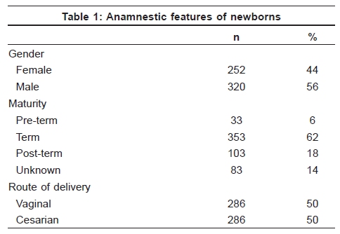
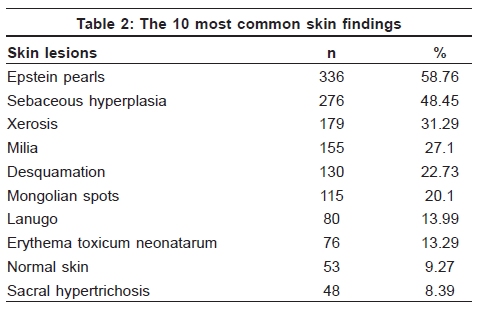
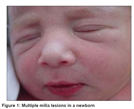
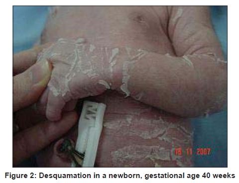
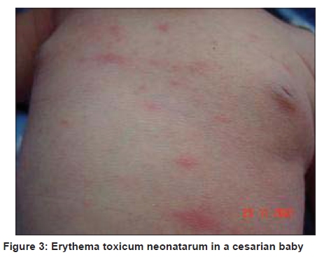
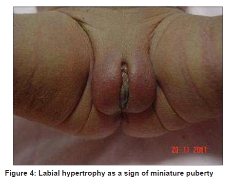
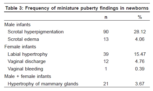
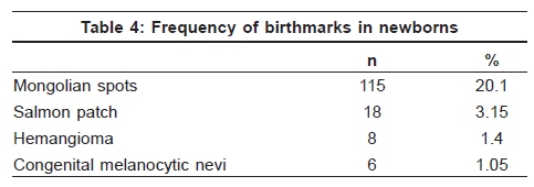
Indian Journal of Dermatology, Venereology and Leprology, Vol. 75, No. 6, November-December, 2009, pp. 638 Net Study Cutaneous lesions in Turkish neonates born in a teaching hospital G. Gokdemir, H. Kaya Erdogan, A. Köslü, B. Baksu1 Departments of Dermatology,
1Third Obstetrics and
Gynecology, Sisli Etfal
Research and Teaching
Hospital, Istanbul, Turkey Code Number: dv09223 PMID: 19915262 DOI: 10.4103/0378-6323.57742 Abstract Background: The neonatal period is regarded as the first 4 weeks of extrauterine life. In the literature, there are numerous articles about the skin findings in neonates and the results of these studies show differences according to races and environmental factors.Aims: Our objective was to evaluate the skin lesions seen in neonates delivered in our hospital and to determine their relationship to gender, gestational age and route of delivery. Methods: Newborns delivered at the Obstetrics Clinics of our hospital between November 2007 and April 2008 were included in this study. Dermatologic examination was performed and relationship between the 10 most common skin findings and gender, gestational age and route of delivery were statistically evaluated. Results: A total of 572 newborns were examined for the presence of skin lesions. Most common skin findings were Epstein pearls (58.76%), sebaceous hyperlasia (48.45%) and xerosis (31.29%). Milia and sebaceous hyperplasia in girls, desquamation and xerosis in preterms, Epstein pearls, sebaceous hyperplasia and desquamation in vaginally delivered babies were found to be more frequent and the differences were statistically significant. Conclusion: We found that 90.7% of the neonates had one or more cutaneous lesions. Maturity and type of delivery of the babies were important factors in their causation. In Turkey, this study is the first study performed on the skin lesions seen during the neonatal period. With this study, we want to increase the awareness about the skin findings in neonates. Keywords: Epstein pearls, Neonatal dermatoses, Newborn Introduction The neonatal period is regarded as the first 4 weeks of extrauterine life. Many cutaneous lesions are seen during this period. The most commonly detected lesions are considered transient as a result of physiologic response and limited to the first several days or weeks of life. Therefore, these conditions are rarely examined by dermatologists. However, it is important to identify them correctly to avoid concerns of parents, gynecologists and pediatricians as well as unnecessary diagnostic or therapeutic procedures. Several studies have been undertaken to assess the frequency of cutaneous lesions in neonates of various countries. [1],[2],[3] This is the first study in the literature from Turkey regarding neonatal dermatoses. The aims of the study were to: (i) assess the prevalence of skin findings in newborns and (ii) estimate the association between gender, gestational age and delivery with skin findings. Methods All consecutive live births at the Gynecology and Obstetrics Clinics in our hospital between November 2007 and April 2008 were recruited into study. Before examination, consent forms were signed by the parents. The gender, gestational age and the mode of delivery were recorded in each case. All neonates were examined by the same dermatologist. The entire skin surface, including the mucous membranes and the nails, was thoroughly examined. The diagnosis of the skin lesions was recorded. No biopsies were performed. Regarding gestational age, three groups (pre-term ≤36 weeks, term 37-42 week, post-term ≥42 week) were established. The information collected for the 10 most common neonatal skin lesions were analyzed using the computarized program SPSS 10 version using the chi-square test. Significance was defined as P< 0.05. Results Five hundred seventy-two newborns were examined during the study period. Of these, 252 (44%) were female and 320 (65%) were male infants. The babies were seen only once between days 1 and 20 (average day: 2.09), the majority (80.9%) being examined within 48 h of birth. The gender, gestational age and mode of delivery of neonates are given in [Table - 1]. Five hundred nineteen neonates (90.7%) had one or more cutaneous findings. The 10 most common neonatal skin lesions are shown in [Table - 2]. Regarding the gender of the neonates, sebaceous hyperplasia and milia [Figure - 1] were more frequent in female infants than in male ones (P< 0.05). For other dermatoses, there was no difference between male and female neonates. Concerning the gestational age, xerosis and desquamation [Figure - 2] were more frequent (P< 0.001) whereas Epstein's pearls and sebaceous hyperplasia were less frequent in post-term babies compared with other groups. With respect to the type of delivery, erythema toxicum neonatarum (ETN) [Figure - 3] was more frequent in the cesarean delivery group (P< 0.05). On the other hand, Epstein's pearls, sebaceous hyperplasia and desquamation were more frequent in the vaginal delivery group than in other groups (P< 0.05). Skin findings of miniature puberty were seen in 176 neonates. Scrotal hyperpigmentation (for only male infants, 28.1%) and labial hypertrophy (for only female infants, 15.4%) [Figure - 4] were the most common findings [Table - 3]. Mongolian spots (20.1%) was the most common lesion among pigmentary abnormalities [Table - 4]. The frequency of Mongolian spots did not vary significantly with gender, gestational age and mode of delivery. The prevalence of congenital melanocytic nevi was 1.05% (six infants). Discussion Several studies about the prevalence of neonatal dermatoses have been documented in various countries and racial groups. Some articles are confined to a particular disease such as melanocytic nevus or limited to a particular time such as first 5 days. [3],[4] In our study, all neonates born in the Obstetrics and Gynecology Clinics were observed, including both transient conditions and birthmarks during the study period. The prevalence of neonatal dermatoses in the literature was reported between 57 and 99.3%. [1],[5],[6] In this present study, most of the neonates (90.7%) had one or more cutaneous findings. These different results may be related to study methods and racial features. Epstein's pearls (58.7%) were the most common finding in our study. We found that Epstein's pearls was less frequent in post-term babies compared with pre-term and term ones. A higher incidence seen in term babies was reported by Moosavi et al. [1] Other studies observed the frequency of Epstein's pearls as 56, 70.2 and 88.7%. [1],[2],[5] Epstein's pearls had not been reported in some studies because the oral mucosa had not been examined in them. It is important to recognize these common lesions because they dissappear spontaneously and they do not need injudicious attempts to be removed. [5],[7] Sebaceous hyperplasia (48.4%) was the second most common finding in our study. The incidence we found is nearly comparable with the incidences observed in other studies. No correlation of sebaceous hyperplasia with gender, racial factors and gestational age was reported. [1],[2],[5] We found that sebaceous hyperplasia was more frequent in pre-term and term babies, but these result might be misleading because of the presence of an unknown maturity group. Xerosis (31.2%) and desquamation (22.7%) were the other common findings in our study. Some of the babies had both skin conditions. In normal term neonates, the skin is smooth and moist at birth. However, benign superficial skin peeling may be seen in infants because of reduction of transepidermal water loss. That term is called as "physiologic desquamation." [7],[8] Xerosis was not mentioned in the other studies. We accepted minimal scaling without clinical desquamation as "xerosis." The incidence of desquamation as observed in the literature varies from 7.2 to 83%. [9] We found that the prevalence of xerosis and desquamation was more common in post-term babies as similar to the study of Moosavi et al. [1] This result is supported with Griffith's hypothesis. However, Rivers et al. found that the prevalence of desquamation did not vary with gestational age. [5] Milia (27.1%) was one of the most commonly observed skin lesions. The prevalence of milia varies from 7.5 to 36%. [1],[2],[5] Our result is nearly comparable with that reported in the literature. We found that it was not correlated with maturity as similar to results of Nanda et al. [2] However, milia was more common in female babies than in male ones. ETN is a common eruption in the neonate, particularly in term infants. Mechanical/termal trauma has been suggested as etiological factors. [7] Contrary to other reports, the incidence of ETN was 13.2%, being more common in cesarian babies in our study. Hence, different factors as genetic, environmental or racial features may play a role in the etiology of ETN. [2],[4],[5],[10] Lanugo hairs and sacral hypertrichosis were the other common findings. The incidence of lanugo is more common in pre-term babies. We found similar results, but it was not statistically significant because of an inadequate number of pre-term babies. [8],[9] Scrotal hyperpigmentation (28.1%) and labial hypertrophy (15.4%) were the most common findings as miniature puberty. Boccardi et al. [11] suggested that hyperpigmentation of the genital area was more common in newborns of non-European origin. It was speculated that the variation in genital hyperpigmentation may be related to the differential activation of melanocytes. Therefore, racial factors and skin type may be important factors. The incidence of labial hypertrophy is nearly comparable with the literature. [11] The prevalence of birthmarks in newborns have been reported in various countries. [3] Mongolian spots is the most common birthmark, as in our study (20.1%). The frequency rate of Mongolian spots showed a marked racial difference. In the literature, various rates (0.1% in Finland, 11.8% in Arabs, 6.65% in Jewish, 62.2% in Indian, 71.3% in Iran, 81.5% in Japan) have been documented. [1],[2],[3],[4],[5] The incidence of congenital melanocytic nevus in our study was 1.05%, which was comparable to other studies. [12] In conclusion, we tried to evaluate the prevalence of skin lesions during the neonatal period and to determine any correlations with gender, gestational age and type of delivery in Turkish newborns born in a teaching hospital. Several skin lesions, physiologic or pathologic, are present at birth and a number of others appear during the neonatal period. It was very important to differentiate the physiologic skin lesions from the pathologic ones. There are many studies with different methods about neonatal dermatoses in the literature from various countries. The frequency of these lesions changes with racial and environmental factors. However, further studies about different factors like maternal history may be useful. Acknowledgment We thank the medical and nursing staff of the Department of Obstetrics and Gynecology in our hospital for their help and cooperation in conducting this study. References
Copyright 2009 - Indian Journal of Dermatology, Venereology and Leprology The following images related to this document are available:Photo images[dv09223t2.jpg] [dv09223f1.jpg] [dv09223t3.jpg] [dv09223f3.jpg] [dv09223f2.jpg] [dv09223t4.jpg] [dv09223t1.jpg] [dv09223f4.jpg] |
| |||||||||

