 |
| About Bioline | All Journals | Testimonials | Membership | News |
|
||||||
|
||||||
Indian Journal of Dermatology, Venereology, and Leprology, Vol. 76, No. 2, March-April, 2010, pp. 159-164 Brief Report Prevalence of different Malassezia species in pityriasis versicolor in central India Rahul Chaudhary, Sanjay Singh, Tuhina Banerjee1 , Ragini Tilak1 Departments of Dermatology and 1Microbiology, Institute of Medical Sciences, Banaras Hindu University, Varanasi-221 005, India Correspondence Address: Dr. Sanjay Singh, C-9, New Medical Enclave, Banaras Hindu University, Varanasi-221 005, India, sanjaye2@gmail.com Code Number: dv10041 PMID: 20228545 DOI: 10.4103/0378-6323.60566 Abstract Background: In the last 10 years, different studies have shown
interesting geographical variations in the prevalence of different Malassezia species
in pityriasis versicolor. Aim: Identification of Malassezia species
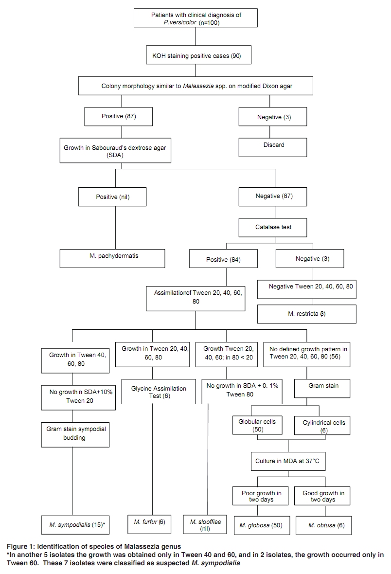
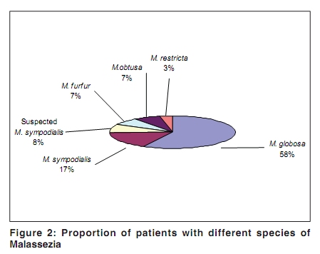
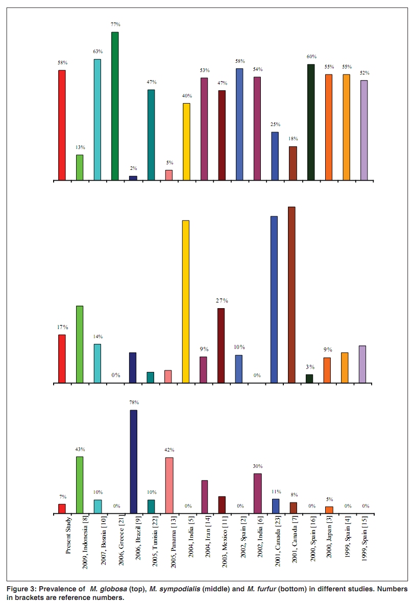
isolated from patients with pityriasis versicolor. Keywords: Malassezia, pityriasis versicolor, Pityrosporum Introduction Malassezia (formerly Pityrosporum) is a genus of related yeasts, naturally found on skin of many animals and humans. Malassezia were originally identified by the French scientist Malassez. Later, they were reclassified into two species: P. ovale, which is lipid-dependent and found only on humans, and P. pachydermatis, which are lipophilic, but not lipid-dependant, and found on most animals. P. ovale was later divided into two classes, P. ovale and P. orbiculare, and later both together were renamed M. furfur. Malassezia is commonly found in seborrheic areas in up to 90% adults and becomes pathogenic under certain circumstances such as warm and humid environment. Currently, Malassezia genus has been enlarged to include 12 species comprising M. furfur, M. pachydermatis, M. sympodialis, M. globosa, M. obtusa, M. restricta, M. slooffiae, M. dermatis, M. japonica, M. nana, M. yamotoensis, and a not formally recognised species M. equi.[1] The first seven species have been well studied in relation to pityriasis versicolor. The most common disease caused by Malassezia is pityriasis versicolor, it has also been implicated in seborrheic dermatitis. In the last 10 years, studies have shown interesting geographical variation in the prevalence of different Malassezia species in pityriasis versicolor. We identified different Malassezia species in pityriasis versicolor and investigated whether the species vary in relation to the color (hypopigmented or hyperpigmented) of lesions. Methods One hundred patients with pityriasis versicolor attending the skin diseases outpatient department of a tertiary care hospital in central India were consecutively included. Skin scrapings were taken from the most scaly site; the study then proceeded as follows to identify the following species: M. furfur, M. pachydermatis, M. sympodialis, M. globosa, M. obtusa, M. restricta, and M. slooffiae Direct Microscopy Morphological characteristics of yeasts were identified in 10% KOH smear and Gram staining. Culture Sabouraud′s dextrose agar (SDA) containing cycloheximide with olive oil overlay and modified Dixon agar (MDA), a more specialized media, which permits better visualisation and isolation of the colonies, were used. Culture plates were examined on days 3, 7 and then at weekly intervals up to three weeks. Biochemical Tests Catalase Reaction Production of gas bubbles on adding a drop of hydrogen peroxide indicated a positive reaction. Assimilation of Glycine as a Nitrogen Source M. furfur is the only species which assimilates glycine. Isolates were inoculated in modified Dixon glycine media (with 7mMol/liter glycine). Plates were incubated at 370C for 3 days. Growth within 2 to 3 days showed glycine assimilation. Negative reporting was done after 7 days. Tween (20, 40, 60, 80) Utilization Tests Plates were systematically incubated for one week at 320C. Utilization of Tweens was assessed by the degree of growth and/or reaction (precipitation) of the lipophilic yeasts around individual wells. Results Of the 100 patients [80 male, 20 female; mean (SD) age, 25.2 (10.6) years], hypopigmented lesions were present in 91 and nine patients had hyperpigmented lesions. Duration of illness ranged from 15 days to 35 years. Forty three patients complained of itching. In 80 patients the lesions were present on back, in 65 on chest, in 62 on neck, in 40 on upper limbs, in 25 on shoulders, and in 23 patients on face. In all patients, lesions were present on multiple sites. In 10 patients, 10% KOH smear was negative, while in 90 patients it showed the characteristic "spaghetti and meatball" appearance. Of these 90 cases, growth was obtained on modified Dixon agar from 87 cases. Fifty isolates (57.5%) were M. globosa, 15 (17.2%) were M. sympodialis, 7 (8.0%) were suspected M. sympodialis, 6 (6.9%) each of the isolates were M. furfur and M. obtusa, and 3 (3.4%) isolates were M. restricta [Figure - 1] and [Figure - 2]. There was no growth in SDA, ruling out the presence of M. pachydermatis, the only lipid-independent species. Other species like M. slooffiae were not isolated. When the frequencies of isolated species were compared between two age groups (=20 versus> 20 years), no significant difference was found for the two most frequently isolated species (x2 test without Yates correction; M. globosa, P=0.32; M. sympodialis, P=0.07). Similarly, there was no significant difference between the genders for the two most common species (M. globosa, P=0.73; M. sympodialis, P=0.74). Of the nine patients with hyperpigmented lesions, M. globosa was isolated in six, M. sympodialis in two and no fungal growth occurred in one case. M. globosa had stable spherical cells on Gram stain. Buds were formed on the narrow base in M. globosa. M. sympodialis had small ovoid cells with sympodial budding, which is a characteristic feature. M. obtusa showed cylindrical cells. M. furfur showed characteristic glycine assimilation. The catalase reaction was positive for all except M. restricta, which is the only lipid-dependent species of Malassezia to consistently lack catalase. The Tween assimilation test allowed the differentiation of most Malassezia species. The growth of 22 isolates was inhibited by high concentrations of Tween 20 in M. sympodialis. Fifty nine isolates did not utilize any of the Tweens (M. globosa, M. obtusa and M. restricta). Discussion Results of our study are most comparable to those of Aspiroz et al,[2] Nakabayashi et al,[3] and Crespo Erchiga et al,[4] who isolated M. globosaat the frequencies of 58.2%, 55%, and 55%, respectively. Two similar studies have been done in India, Kindo et al,[5]showed that in south India M. sympodialis is the commonest agent (58.3%) followed by M. globosa (39.6%). Another study from north-central India, conducted by Dutta et al,[6] showed that 54% of isolates belonged to M. globosa, the next common species was M. furfur (30%). In the present study, we found a group in which all results were consistent with M. sympodialis i.e. KOH positivity, glistening, smooth, flat colony on MDA, and sympodial budding; but the Tween test showed positivity either only on Tween 40 and 60, or only on Tween 60, instead of showing positivity in Tween 40, 60, and 80. We called this group suspected M. sympodialis, of which we found 7 isolates. Of the previous 17 studies, 11 showed M. globosa to be the most common species isolated. Its pathogenicity might be explained by high lipolytic activity.[2]A few studies have found M. sympodialisto be the most common species;[5],[7]we found it to be second most frequent, as in other studies also.[3],[8],[9],[10],[11] M. furfur is also responsible for pityriasis versicolor, particularly under tropical climate.[12]In the present study, this species was isolated in 6.9% of patients, quite similar to another report (7.8%).[7]Some studies have found much higher prevalence of M. furfur,[8],[9],[13]isolated as the most common agent. M. obtusawas isolated in 6.9% of patients in the present study, similar to a previous report (8.1%).[14]We found no isolates of M. slooffiae. In the present study, M. restrictawas isolated in 3.4% of cases, similar to a previous report (3%).[9]In contrast to a previous study,[15]we found no isolate of M. pachydermatis. Many other studies have also not found this species, indicating that this species may not be considered the causal organism for pityriasis versicolor. The comparisons among results of different studies have been summarized in [Figure - 3]. Because the number of patients with hyperpigmented lesions of pityriasis versicolorwas only nine, no definite conclusions could be drawn about the relationship of species and the pigmentory changes produced. As the predominant isolate in both hyperpigmented and hypopigmented lesions was M. globosa, probably no such relationship exists. The differences in frequencies of Malassezia species among different studies may be attributed to different culture media (modified Dixon agar/Leeming-Notman agar) and perhaps to ethnic and geographic factors.?[16]The identification of Malassezia yeast to species level is of importance to determine which species are implicated in certain skin disease and whether there is variation in the distribution of the yeast with clinical data, body site, origin of the population etc. Further, the results of the in vitrosusceptibility studies have shown variations in susceptibility of the seven Malassezia species to various antifungal agents. Strains of M. furfur, M. globosa and M. obtusa have been found to be more tolerant to terbinafine than the remaining species, while M. sympodialiswas highly susceptible.[17]These results suggest that correct identification of Malassezia species may be important for the selection of appropriate antifungal therapy. Molecular methods such as nested-PCR[18],[19] or PCR-REA[20]are being developed to solve the problems arising due to time-consuming morphological and physiological techniques and the difficulty in interpretation of some physiological patterns. Further studies will help determine whether the species variation found in different studies are related to ethnic differences or to climatic influence or the site or type (hypo- or hyperpigmented) of lesions.[23] References
Copyright 2010 - Indian Journal of Dermatology, Venereology, and Leprology The following images related to this document are available:Photo images[dv10041f2.jpg] [dv10041f3.jpg] [dv10041f1.jpg] |
| |||||||||

