 |
| About Bioline | All Journals | Testimonials | Membership | News |
|
||||||
|
||||||
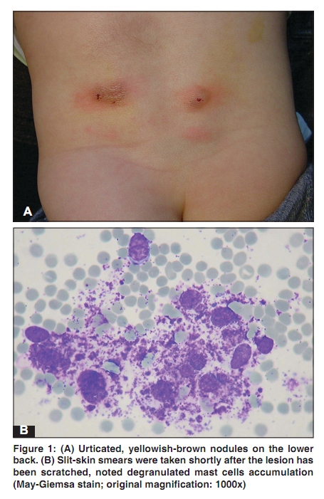
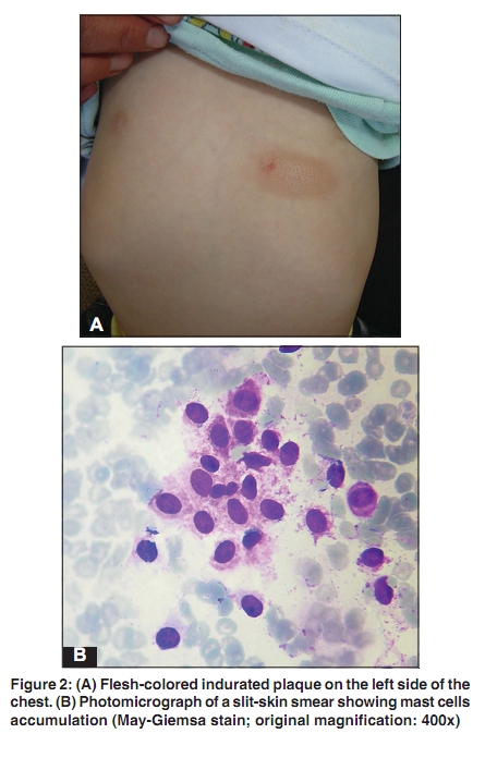
Indian Journal of Dermatology, Venereology, and Leprology, Vol. 76, No. 2, March-April, 2010, pp. 187-189 Letter to the Editor Slit-skin smear in diagnosis of cutaneous mastocytomas Mehmet Harman, Sedat Akdeniz, Bülent Mizrak1 Departments of Dermatology and 1Pathology, Faculty of Medicine, Dicle University, Diyarbakır, Turkey Correspondence Address: Dr. Mehmet Harman, Dicle Üniversitesi, Tıp Fakültesi, Dermatoloji Anabilim Dalı, 21280 Diyarbakır, Turkey, mharman@dicle.edu.tr Code Number: dv10051 PMID: 20228555 DOI: 10.4103/0378-6323.60559 Sir, Cutaneous mastocytosis occurs relatively commonly in infants and children. It presents clinically in several distinct forms. Large mast cell aggregates in the form of solitary or multiple mastocytomas are observed most frequently, and arise most commonly in infants. The diagnosis of mastocytosis is made on the basis of history and clinical presentation and is confirmed by histopathologic examination of the lesional skin specimen obtained by a skin biopsy. [1] We report here two illustrative cases of cutaneous mastocytoma diagnosed with slit-skin smear examination and discuss the advantages of the slit-skin smear test in the diagnosis of mastocytoma. The first case was a 6-month-old male infant presented with slowly growing asymptomatic nodular growths on his back since birth. Clinical examination revealed yellowish-brown, round-to-oval, multiple nodules. Inflammatory flare characterized with redness and swelling was observed in the lesions after scratching [Figure - 1]A. Systemic examination was unremarkable, and routine laboratory tests were within normal limits. Slit-skin smear specimens were obtained from the most remarkable lesion. After cleaning with the alcohol, the lesion was grasped between the thumb and forefinger of the non-dominant hand until the site was blanched. About 5 mm long and 3 mm deep incision was made with Bard Parker No 15 blade and scraped inside the cut with side of the scalpel to obtain tissue and pulp. Microscopic examination of the slit skin smears stained with Giemsa stain showed degranulated mast cell accumulations [Figure - 1]B. The second case was an 11-month-old male infant presented with a slowly growing plaque on his chest that had been present since birth. Clinical examination revealed solitary, flesh-colored, measuring 3 ´ 2 cm in size, indurated oval plaque on the left side of his chest [Figure - 2]A. An urticarial attack associated with transient redness and swelling in the lesion 3 days earlier was reported by his mother. His systemic examination was unremarkable, and routine laboratory tests were within normal limits. Slit-skin smear specimens were obtained from the lesion. Microscopic examination of the slit skin smears stained with Giemsa stain showed mast cell accumulations [Figure - 2]B. Based on cytologic findings and clinical data of both cases, the definitive diagnosis of mastocytoma was made. Mastocytosis is a group of disorders characterized by mast cell proliferation and accumulation within various organs, most commonly in the skin. The term mastocytoma has been used to describe nodular infiltrates of mast cells occurring as a single or several isolated lesions. Clinically, it is characterized with a single or several, red to reddish-brown minimally infiltrated nodule or plaque. Darier′s sign is present in the majority of patients with cutaneous mastocytosis. [1],[2] Recognition of mastocytoma can be difficult, especially in patients who do not have the characteristic skin lesions and Darier′s sign. The clinical differential diagnosis includes congenital nevus, juvenile xanthogranuloma, xanthoma, spitz nevus, insect bite reaction, and nodular scabies. Definitive diagnosis of mastocytoma requires histopathologic examination of the lesional skin specimen obtained by a skin biopsy. Diagnostic cytology is a relatively new science. As a method for the diagnosis of cutaneous disorders, cytology was first used by Arnoult Tzanck in 1947, for the diagnosis of vesiculo-bullous disorders.[3] Since then, cytology has been widely used by dermatologists in the diagnosis of various cutaneous dermatoses. [4],[5] A definitive diagnosis of mastocytoma can be made by slit-skin smears stained with toluidine blue or Giemsa stain used to identify mast cells that are characterized by the presence of metachromatic granules in their cytoplasm. Slit-skin smear in infants and small children can be performed successfully, safely, and easily. In this cytodiagnostic technique, a sample of material is collected from a tiny cut in the lesional skin, placed on a glass slide, stained, and then examined under microscope. Local anesthesia is not required. We suggest that slit-skin smear examination in clinically suspected cases of mastocytoma may prove to be a rapid, non-invasive technique to support the diagnosis. References
Copyright 2010 - Indian Journal of Dermatology, Venereology, and Leprology The following images related to this document are available:Photo images[dv10051f1.jpg] [dv10051f2.jpg] |
| |||||||||

