
|
Indian Journal of Dermatology, Venereology and Leprology
Medknow Publications on behalf of The Indian Association of Dermatologists, Venereologists and Leprologists (IADVL)
ISSN: 0378-6323 EISSN: 0973-3922
Vol. 76, Num. 2, 2010, pp. 206-212
|
Indian Journal of Dermatology, Venereology, and Leprology, Vol. 76, No. 2, March-April, 2010, pp. 206-212
Resident's Page
Named bodies in dermatology
Fiona F. Sequeira, Ambika Kumar, Usha Kini1, Jayanthi2
Departments of Dermatology, 1Pathology, 2Microbiology, St John’s Medical College and Hospital, Karnataka, India
Correspondence Address: Dr. Fiona F Sequeira, Department of Dermatology,
St John’s
Medical College and Hospital, Koramangala, Bangalore, Karnataka - 560 034,
India, dr_fiona@rediffmail.com
Code Number: dv10060
PMID: 20228564
DOI: 10.4103/0378-6323.60551
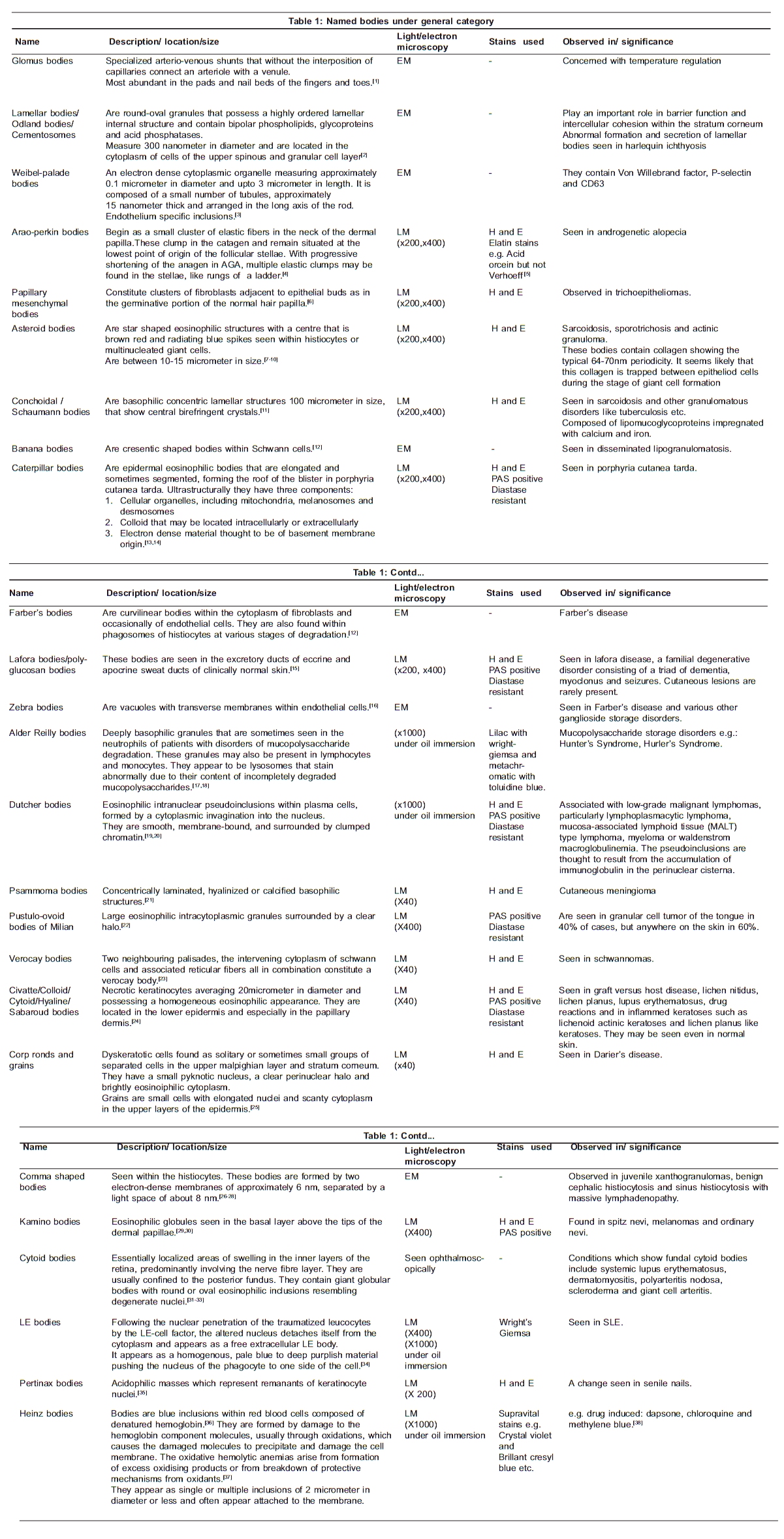
Introduction This article cites, briefly and completely, the named bodies in dermatology. It is not restricted to bodies seen only histopathologically, rather an attempt has been made to include bodies found in the skin, blood smears, fundal examination, lymph nodes, culture media and bone marrow smears provided they pertain to disease conditions of our subject of interest [Figure - 1],[Figure - 2],[Figure - 3],[Figure - 4]. At the end of the day, the article will stand useful to not only the dermatologist / pathologist but more so to the young budding dermatology/ pathology postgraduates who are often quizzed on this part of dermatology. The article has been broadly divided into the following categories: General Category [Table - 1]
- Normal cutaneous anatomy: Glomus bodies, lamellar bodies, Weibel-palade bodies
- Hair disorders: Arao-Perkin bodies, papillary mesenchymal bodies
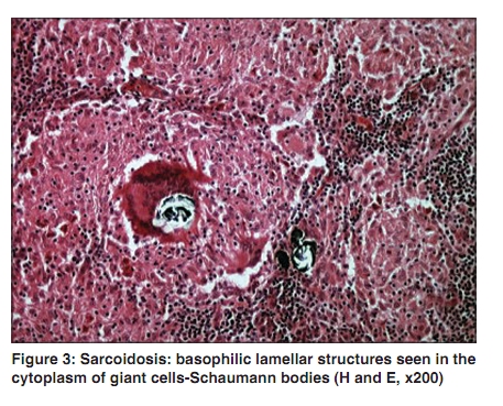
- Granulomatous disorders: Asteroid bodies, Conchoidal /Schaumann bodies
- Metabolic and storage disorders: Banana bodies, Caterpillar bodies, Farber′s bodies, Lafora bodies, Zebra bodies, Alder Reilly bodies
- Tumors: Dutcher bodies, Psammoma bodies, Pustulo-ovoid bodies of Milian, Verocay bodies
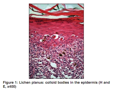
- Papulosquamous disorders: Civatte bodies, Corp ronds and grains
- Histiocytic disorders: Comma shaped bodies
- Benign pigmented lesions: Kamino bodies
- Collagen vascular disorders: Cytoid bodies, LE bodies
- Age related change: Pertinax bodies
- Drug induced: Heinz bodie
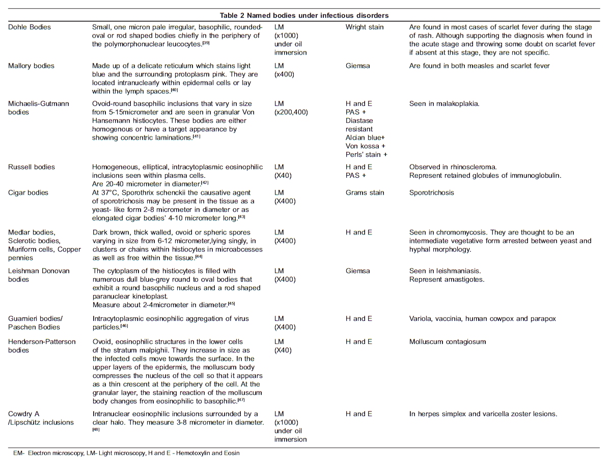
Infectious Disorders [Table - 2]
- Bacterial: Dohle bodies, Mallory bodies, Michaelis-Gutmann bodies, Russell bodies, Donovan bodies, Gamna favre bodies
- Fungal: Cigar bodies, Medlar bodies
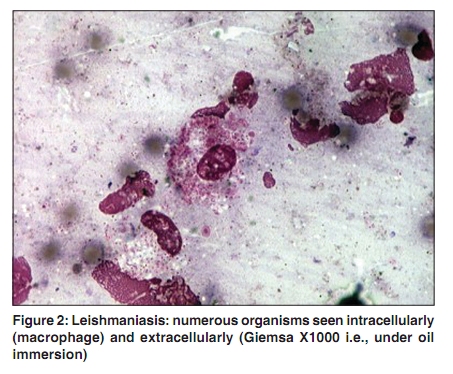
- Protozoal: Leishman Donovan bodies
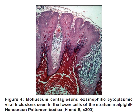
- Viral: Poxviridae: Guarnieri bodies, Henderson-Patterson bodies:
- Herpes group: Cowdry A.
[50]
References
| 1. | Murphy GF. Histology of the skin. In: Elder DE, Elenitsas R, Johnson B, Murphy G, editors. Lever's histopathology of the skin. 9th ed. Philadelphia:JB Lippincott; 2005. p. 9-58. Back to cited text no. 1 |
| 2. | Jakubovic HR, Ackerman AB. Structure and function of the skin. In: Moschella SL, Hurley HJ, editors. Moschella's Dermatology.2nd ed. Philadelphia: WB Saunders Co; 1992. p. 14-5. Back to cited text no. 2 |
| 3. | Thorgeirsson G, Robertson AL Jr. The vascular endothelium pathobiologic significance. Am J Pathol 1978;93:803-48. Back to cited text no. 3 |
| 4. | Nagle RB, Witte MH, Martinez AP, Witte CL, Hendrix MJ, Way D, et al. Factor VIII- associated antigen in human lymphatic endothelium. Lymphology 1987;20:20-4. Back to cited text no. 4 |
| 5. | Pinkus H. Differential patterns of elastic fibres in scarring and non-scarring alopecias. J Cutan Pathol 1978;5:93-104. Back to cited text no. 5 |
| 6. | Rapini RP. Follicular neoplasms. In: Rapini RP,editor. Practical Dermatopathology.1st ed. China: Mosby; 2005. p. 286. Back to cited text no. 6 |
| 7. | Lever WF, Freiman DG. Sarcoidosis:a report of a case with erythrodermic lesions, subcutaneous nodes and asteroid inclusion bodies in giant cells. Arch Derm Syphilol 1948;57:639-54. Back to cited text no. 7 |
| 8. | Gawkrodger DJ. Sarcoidosis. In:Burns T, Breathnach S, Cox N, Griffiths C, editors. Rook's Textbook of dermatology. 7th ed. Blackwell Science: Oxford; 2004. p. 58-4. Back to cited text no. 8 |
| 9. | Brien JP. Actinic granuloma:the expanding significance. Int J Dermatol 1985;24:473-90. Back to cited text no. 9 |
| 10. | Azar HA, Lunardelli C. Collagen nature of asteroid bodies of giant cells in sarcoidosis. Am J Pathol 1969;57:81-92. Back to cited text no. 10 |
| 11. | Winkelmann RK, Dahl PR, Perniciaro C. Asteroid bodies and other cytoplasmic inclusions in necrobiotic xanthogranuloma with paraproteinemia. J Am Acad Dermatol 1998;38:967-70. Back to cited text no. 11 |
| 12. | Rauch HJ, Auböck L. "Banana bodies" in disseminated lipogranulomatosis (farber's disease). Am J Dermatopathol 1983;5:263-6. Back to cited text no. 12 |
| 13. | Egbert BM, LeBoit PE, McCalmont T, Hu CH, Austin C. Caterpillar bodies: distinctive, basement membrane containing structures in blisters of porphyria. Am J Dermatopathol 1993;15:199-202. Back to cited text no. 13 |
| 14. | Raso DS, Greene WB, Maize JC, McGown ST, Metcalf JS. Caterpillar bodies of porphyria cutanea tarda ultrastructurally represent a unique arrangement of colloid and basement membrane bodies. Am J Dermatopathol 1996;18:24-9. Back to cited text no. 14 |
| 15. | Karimipour D, Lowe L, Blaivas M, Sachs D, Johnson TM. Lafora disease: Diagnosis by skin biopsy. J Am Acad Dermatol 1999;41:790-79. Back to cited text no. 15 |
| 16. | Dolman CL.Diagnosis of neurometabolic disorders by examination of skin biopsies and lymphocytes. Semin diagn pathol 1984;1:82-97. Back to cited text no. 16 |
| 17. | Alder A. Ueber konstitutionell bedingte Granulationsveraenderungen der Leukocyten. Dtsch Arch Klin Med 1939;183:372-8. Back to cited text no. 17 |
| 18. | Reilly WA. The granules in the leukocytes in gargoylism. Am J Dis Child 1941;62:489-491. Back to cited text no. 18 |
| 19. | Dutcher TF, Fahey JL. The histopathology of the macroglobulinemia of Waldenstrom. J Natl Cancer Inst 1959;22:887-917. Back to cited text no. 19 |
| 20. | Brunning RD, Parkin J. Intranuclear inclusions in plasma cells and lymphocytes from patients with monoclonal gammopathies. Am J Clin Pathol 1976;66:10-21. Back to cited text no. 20 |
| 21. | Rapini RP. Neural neoplasms. In: Rapini RP,editor. Practical Dermatopathology. 1 st ed. China: Mosby; 2005. p. 338. Back to cited text no. 21 |
| 22. | Rapini RP. Neural neoplasms. In: Rapini RP, editor. Practical Dermatopathology. 1 st ed. China: Mosby; 2005. p. 336. Back to cited text no. 22 |
| 23. | Reed R, Argenyi Z. Tumors of neural tissue. In: Elder DE, Elenitsas R, Johnson B, Murphy G, editors. Lever's histopathology of the skin. 9th ed. Philadelphia: JB Lippincott, 2005. p. 1116-22. Back to cited text no. 23 |
| 24. | Grubauer G, Romani N, Kofler H, Stanzl U, Fritsch P, Hintner H.Apoptotic keratin bodies as autoantigens causing the production of IgM and antikeratin intermediate filament autoantibodies. J Invest Dermatol 1986;87:466. Back to cited text no. 24 |
| 25. | Stefen C. Dsykeratosis and the dyskeratoses. Am J Dermatopathol 1988;10:356-63. Back to cited text no. 25 |
| 26. | Török E, Daróczy J. Juvenile xanthogranuloma: An analysis of 45 cases by clinical follow-up, light- and electron microscopy. Acta Derm Venereol 1985;65:167-9. Back to cited text no. 26 |
| 27. | Eisenberg EL, Bronson DM, Barsky S. Benign cephalic histiocytosis. A case report and ultrastructural study. J Am Acad Dermatol 1985;12:328-31. Back to cited text no. 27 |
| 28. | Avril MF, Amiel JL, Théodore C, Chahine G, Caillaud JM, Caillou B, et al. Manifestations cutanées du syndrome de Rosai et Dorfman. Ann Dermatol Venereol 1984;111:661-2. Back to cited text no. 28 |
| 29. | Arbuckle S, Weedon D. Eosinophilic globules in the spitz nevus. J Am Acad Dermatol 1982;7:324-7. Back to cited text no. 29 |
| 30. | Kamino H, Flotte TJ, Misheloff E, Greco MA, Ackerman AB. Eosinophilic globules in spitz's nevi. New findings and a diagnostic sign. Am J Dermatopathol 1979;1:319-24. Back to cited text no. 30 |
| 31. | Ashton N. Pathophysiology of retinal cotton-wool spots. Br Med Bull 1970;26:143-50. Back to cited text no. 31 |
| 32. | Arroyo JG. Cotton-Wool Spots May Challenge Diagnosis. Rev Ophthalmol 2004;11:4. Back to cited text no. 32 |
| 33. | Gold DH, Morris DA, Henkind P. Ocular findings in systemic lupus erythematosus. Br J Ophthalmol 1972;56:800-4. Back to cited text no. 33 |
| 34. | Robineaux R, Pinet J. Hargraves' cell. Description, significance and research technique. Rev Prat 1965;15:2523-30. Back to cited text no. 34 |
| 35. | Cohen PR, Scher RK. Aging. In: Hordinsky MK, Sawaya ME, Scher RK,editors. Atlas of hair and nails. Philadelphia: Churchill Livingstone; 2000. p. 213-25. Back to cited text no. 35 |
| 36. | Jacob H, Winterhalter K. Unstable Hemoglobins: The Role of Heme Loss in Heinz Body Formation. Proc Natl Acad Sci U S A 1970;64:697-701. Back to cited text no. 36 |
| 37. | Winterbourn CC, Carrell RW. Studies of Hemoglobin Denaturation and Heinz Body Formation in the Unstable Hemoglobins. J Clin Invest 1974;54:678-89. Back to cited text no. 37 |
| 38. | Goldstein BD. Exacerbation of dapsone-induced Heinz body hemolytic anemia following treatment with methylene blue. Am J Med Sci 1974;267:291-7. Back to cited text no. 38 |
| 39. | Place EH. Scarlet fever. Am J Public Health 1904:767-76. Back to cited text no. 39 |
| 40. | Field CW. On the presence of certain bodies in the skin and blister fluid from scarlet fever and measles. J Exp Med 1905;7:343-50. Back to cited text no. 40 |
| 41. | Palou J, Torras H, Baradad M, Bombí JA, Martín E, Mascaró JM. Cutaneous malakoplakia. Report of a case. Dermatologica 1988;176:288-92. Back to cited text no. 41 |
| 42. | Lucas S. Bacterial Diseases. In: Elder DE, Elenitsas R, Johnson B, Murphy G, editors.Lever's histopathology of the skin. 9th ed. Philadelphia: JB Lippincott; 2005. p. 581. Back to cited text no. 42 |
| 43. | Maberry JD, Mullins JF, Stone OJ.Sporotrichosis with demonstration of hypae in human tissue. Arch Dermatol 1966;93:65-7. Back to cited text no. 43 |
| 44. | McGinnis MR. Chromoblastomycosis and phaeohyphomycosis: new concepts, diagnosis and mycology. J Am Acad Dermatol 1983;8:1-16. Back to cited text no. 44 |
| 45. | Sellheyer K, Haneke E. Protozoan diseases and parasitic infestations. In: Elder DE, Elenitsas R, Johnson B, Murphy G, editors. Lever's histopathology of the skin. 9th ed. Philadelphia: JB Lippincott; 2005. p. 635-40. Back to cited text no. 45 |
| 46. | Xu X, Erickson LA, Elder DE. Diseases caused by viruses. In: Elder DE, Elenitsas R, Johnson B, Murphy G, editors. Lever's histopathology of the skin.9th ed. Philadelphia: JB Lippincott; 2005. p. 660-1. Back to cited text no. 46 |
| 47. | Xu X, Erickson LA, Elder DE. Diseases caused by viruses. In: Elder DE, Elenitsas R, Johnson B, Murphy G, editors .Lever's histopathology of the skin.9th ed. Philadelphia: JB Lippincott; 2005. p. 662-3. Back to cited text no. 47 |
| 48. | Xu X, Erickson LA, Elder DE. Diseases caused by viruses. In: Elder DE, Elenitsas R, Johnson B, Murphy G, editors .Lever's histopathology of the skin. 9th ed. Philadelphia: JB Lippincott; 2005. p. 652-7. Back to cited text no. 48 |
| 49. | Weedon D.Bacterial and rickettsial infections. In: Weedon D,editor. Skin pathology. 1st ed. Edinburgh: Churchill Livingstone; 1997. p. 633-4. Back to cited text no. 49 |
| 50. | Stary A. Sexually transmitted diseases. In: Bolognia JL, Jorizzo JL, Rapini RP, editors. Dermatology, 1st ed. Philadelphia: Mosby; 2003. p. 1291. Back to cited text no. 50 |
Copyright 2010 - Indian Journal of Dermatology, Venereology, and Leprology
The following images related to this document are available:
Photo images
[dv10060t2.jpg]
[dv10060f2.jpg]
[dv10060f1.jpg]
[dv10060t1.jpg]
[dv10060f3.jpg]
[dv10060f4.jpg]
|
