 |
| About Bioline | All Journals | Testimonials | Membership | News |
|
||||||
|
||||||
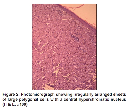
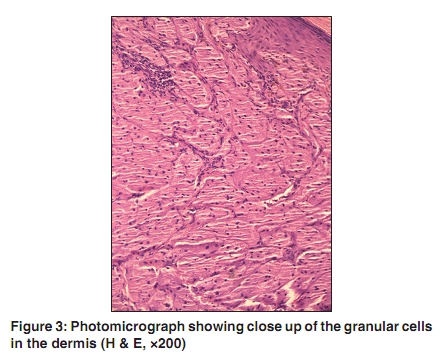
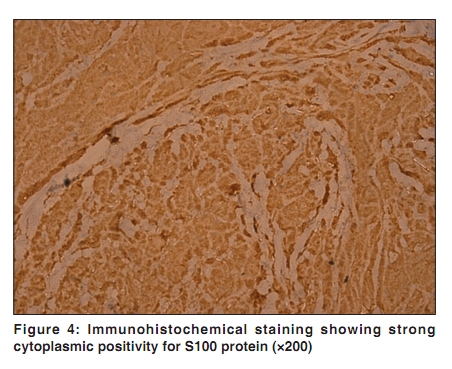
Indian Journal of Dermatology, Venereology, and Leprology, Vol. 76, No. 3, May-June, 2010, pp. 263-265 Case Report Giant granular cell tumor of the vulva Vandana Mehta, C. Balachandran, Laxmi Rao1 , V. Geeta1 Departments of Skin & STD, 1Pathology, Kasturba Medical College, Manipal, Karnataka, India Correspondence Address: Dr. Vandana Mehta, Department of Skin & STD, Kasturba Medical College, Manipal - 576 104, Karnataka, India, vandanamht@yahoo.com Code Number: dv10080 PMID: 20445297 DOI: 10.4103/0378-6323.62966 Abstract A 55-year-old lady presented with a large skin colored growth on her vulva since the age of 15 years, which gradually increased to the present state, with the development of a new lesion on her left thigh. There were no systemic symptoms. Biopsy followed by immunohistochemistry showed features consistent with a granular cell tumor.Keywords: Granular cell tumor, vulva, large Introduction Granular cell tumor (GCT) is a rare benign tumor of debatable histogenesis. It was first described by Abrikossoff in 1926, who postulated a myogenic origin and termed it as granular cell myoblastoma. [1] GCT may occur in almost any part of the body with majority of the tumors occurring in the head and neck region (45-65%). [2] We report a case of a large GCT on the vulva of a healthy female. Case Report A 55-year-old healthy lady presented with a large, skin colored growth on her right labium majus that had been present since the age of 15 years. It was small at onset and had not bothered her until five months back when she noticed a sudden increase in the size of the lesion as well as developed a new lesion on the adjoining left thigh. There was no history of any systemic symptoms. She reported itching over the lesion with development of a few superficial erosions over the large nodule. Cutaneous examination revealed a large skin colored nodular mass measuring 7x5x5 cms on her right labium majus. It was firm to hard in consistency and was formed by a confluence of multiple small papulonodules. Overlying it a few superficial erosions were seen. On the left thigh a similar nodule was present measuring 1x1 cms [Figure - 1]. There was no lymphadenopathy. A biopsy for histopathology from the tumoral mass over right labia majus and the nodule on the thigh revealed irregularly arranged sheets and nests of large polygonal cells with a small hyperchromatic nucleus and abundant coarse eosinophilic granular cytoplasm [Figure - 2] and[Figure - 3]. The cytoplasmic granules were periodic acid schiff (PAS) positive and diastase resistant. Immunohistochemical staining revealed strong cytoplasmic expression of S100, suggesting a diagnosis of granular cell tumor [Figure - 4]. Patient was advised wide surgical excision of the lesion. Discussion Granular cell tumor (GCT) is a rare benign tumor of neurogenic origin resembling Schwann cells. Before the era of electron microscopic and immunohistochemical studies, GCTs were known by a myriad of names such as granular cell myoblastoma, granular cell neurofibroma, granular cell schwannoma, Abrikossoff's tumor and myoblastic myoma. Although GCT may occur in patients of all age groups, they are most frequently encountered between the third to sixth decades of life. Black females are twice as commonly affected than their Caucasian counterparts. Clinically, GCT presents as a solitary, skin colored, asymptomatic nodule less than 3 cms in diameter. In 3-10% of cases there may be multiple lesions. Pruritus and pain have been reported occasionally. Larger lesions may sometimes show surface ulceration, which may clinically give an impression of a malignant neoplasm. GCTs are usually benign; however, malignant transformation may occur in 1-2% cases. Apart from histology, tumor size greater than 5 cms, vascular invasion, necrosis, and rapid growth are important indicators of malignant behavior. [3] Histologically, granular cell tumor is diagnosed by the presence of a characteristic granular cell, which is a large pale polyhedral cell with abundant fine or coarsely granular eosinophilic cytoplasm and a pale staining nucleus situated centrally. The main morphologic feature is the granularity of the cytoplasm which is caused by the massive accumulation of phagolysosomes. Immunohistochemical stains are positive for S100 protein, neuron specific enolase, peripheral nerve myelin proteins and vimentin. [4] Granular cell tumors mostly follow a benign course but, since they are locally infiltrative, most authors recommend complete surgical excision with uninvolved margins to prevent clinical recurrence. Clinically the differential diagnosis should include benign cystic lesions such as bartholin gland tumors, sebaceous cysts and other nodular benign painless vulvar lesions such as dermatofibromas, hidradenomas, lipomas and papillomas. GCT on the vulva is uncommon and vulvar involvement has been reported in 5-16% cases. [5] Laxmisha et al. reported a case of GCT on the clitoris of an 18-year-old female, which was excised; subsequently, with no recurrence at six months follow-up. [6] Ours is probably the second such case. Our case is interesting because she had two GCTs, and one of them was very large in size than what has been conventionally reported in literature. Hence we chose to word it as a "giant granular cell tumor". She was advised regarding wide surgical excision of the tumoral mass. References
Copyright 2010 - Indian Journal of Dermatology, Venereology, and Leprology The following images related to this document are available:Photo images[dv10080f4.jpg] [dv10080f3.jpg] [dv10080f2.jpg] [dv10080f1.jpg] |
| |||||||||
