 |
| About Bioline | All Journals | Testimonials | Membership | News |
|
||||||
|
||||||
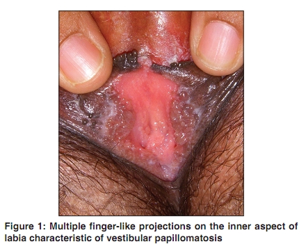
Indian Journal of Dermatology, Venereology, and Leprology, Vol. 76, No. 3, May-June, 2010, pp. 270-272 Case Report Vulvar vestibular papillomatosis U. Wollina, Shyam Verma Department of Dermatology and Allergology, Hospital Dresden-Friedrichstadt, Friedrichstrasse 41, 01067, Germany and Vadodara, India Correspondence Address: Dr. Shyam Verma, 18 Amee Society, Diwalipura, Next to Rajnigandha Apartments, Vadodara, India, vermaderma@rediffmail.com Code Number: dv10082 PMID: 20445299 DOI: 10.4103/0378-6323.62971 Abstract Vulvar vestibular papillomatosis is considered an anatomical variant of the vulva. Recognition of this condition enables one to distinguish it from warts and therefore avoid unnecessary therapy. A 29-year-old lady presented to this clinic with a history of 'small growths' in her vulva since two years. Examination identified skin colored translucent papules; some of them appeared digitate and were seen on the vestibule and inner aspect of both labia minora. They were soft to feel and non-tender. Few lesions looked like elongated pearly penile papules. A provisional diagnosis of vestibular papillomatosis was made and a biopsy was done. It showed finger-like protrusions of loosely arranged subdermal tissue with blood vessels and which were covered by normal mucosal epithelium. No koilocytes were seen and the diagnosis of vestibular papillomatosis was confirmed. We believe that this is the first case report of vulvar vestibular papillomatosis in Indian dermatologic literature.Keywords: First report, India, vulvar vestibular papillomatosis Introduction Vulvar vestibular papillomatosis is an anatomical variant of the vulva. [1],[2] In a prevalence study in London, UK, one per cent of women showed vestibular papillomatosis. [3] Other authors found prevalence rates between 5.1 to 33%. [4] From our experience with a multidisciplinary vulva clinic, the rate seems to be around 5% (unpublished observation). Due to its 'wart like' appearance, it is sometimes misdiagnosed as genital warts. This often leads to unwarranted and aggressive investigations and therapy. Therefore, dermatologists should be familiar with this condition. Vulvar vestibular papillomatosis can be considered the female equivalent of pearly penile papules. [4] Case Report A 29-year-old lady presented to this clinic with a history of 'small growths' in her vulva for two years. She had noticed multiple finger like elongated lesions on the vulva that were gradually increasing in size for the past two years. She also experienced pain during sex. She appeared to be very anxious and feared that the growths might be cancerous and therefore wanted treatment. She had seen two gynecologists and two dermatologists before attending our clinic and had treatments with podophyllin and cryosurgery on three occasions without any improvement of the condition. Our patient was a healthy married nulliparous woman. She had a monogamous relationship with her husband with no history of any extramarital sexual contacts since marriage, four years ago. She also complained of an occasional thick white discharge, associated with recurrent itching. Examination identified a normal vulva except for the lesions she was complaining about. There were no vulval or vaginal ulcers. Examination identified skin colored translucent, papules some of which appeared digitate and were seen on the vestibule and inner aspect of both labia minora [Figure - 1]. They were soft to feel and non-tender. Few lesions looked like elongated pearly penile papules. Some angiokeratomas were also seen on the labia adjacent to these lesions. A provisional diagnosis of vulvar vestibular papillomatosis was made and a biopsy was done. It showed finger-like protrusions of loosely arranged subdermal tissue with blood vessels, which were covered by normal mucosal epithelium [Figure - 2]. No koilocytes were seen and the diagnosis of vulvar vestibular papillomatosis was confirmed. The patient was reassured about the benign nature of the disease, that there was no evidence of infection or malignancy and that no treatment was required. Discussion Vestibular papillomatosis is an uncommon benign condition, which we believe, has not been described in Indian dermatologic literature. Prevalence rates of 1% have been described in literature. [3] Until now less than 20 reports have been published in the international literature since the first description of this condition in 1981 by Altmeyer et al, from Germany. [1],[2] They named the lesions "pseudocondylomata" but it is also known by various other names like 'hirsuties papillaris vulvae', 'hirsutoid papillomas of vulvae', 'vestibular microwarts', 'micropapillomatosis', and 'vulval squamous papillomatosis'. [1],[2],[3],[4] It is considered to be a variant of normal vulvar anatomy with no known significant associations. [1],[2],[3],[4],[5],[6],[7] Vestibular papillomatosis has been recorded in healthy young women in the range of 1 to 33%. [3],[4] The papillae of 1 to 2 mm diameter have the same color as the adjacent mucosa The lesions are soft and are symmetrical or may be linear. [1],[2],[3],[4] They may cover labia minora and the introitus vaginae to a variable extent. [3],[4] They may resemble warts but are distinguished by the fact that the bases of individual papules remain separate unlike in warts where filiform projections tend to fuse at the base and lesions are not confined to the vestibule or the inner aspects of labia minora. [4],[5],[6] In addition, application of 5% acetic acid causes whitening of the lesions in warts whereas vestibular papillae remain unchanged. [3],[4],[5] Human papillomavirus (HPV) infection is not a cause of vestibular papillomatosis as shown by HPV polymerase chain reaction (PCR) or in situ hybridization. [7],[8],[9],[10],[11] Some authors suggest that vestibular papillomatosis, though not caused by HPV infection, could possess a risk of further genital HPV infection, but no data from prospective trials substantiate such a view. [3] The histology of this condition is characterized by finger-like protrusions of a loose connective tissue covered by normal vulvar epithelium. Some vacuolated epithelial cells can occur. This can be explained on the basis that the vestibule comprises of very heavily glycogenated epithelial cells which when subjected to tissue processing get vacuolated and therefore may resemble koilocytes seen with viral infection. [1],[2],[3] In conclusion, vestibular papillae are a normal anatomical variant of the vulva and may be considered as the female equivalent of pearly penile papules in men. Diagnostic characteristics have been summarized by Moyal-Barraco et al [Table - 1]. [12] Following this line, misdiagnosis as vulval warts and unnecessary treatment causing much anguish to the patient can be avoided. Vestibular papillae can be an added source of anxiety for an already anxious patient presenting with associated vulvodynia as it was the case in the present patient. References
Copyright 2010 - Indian Journal of Dermatology, Venereology, and Leprology The following images related to this document are available:Photo images[dv10082t1.jpg] [dv10082f1.jpg] [dv10082f2.jpg] |
| |||||||||
