 |
| About Bioline | All Journals | Testimonials | Membership | News |
|
||||||
|
||||||
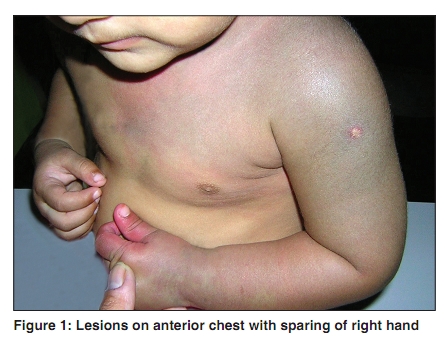
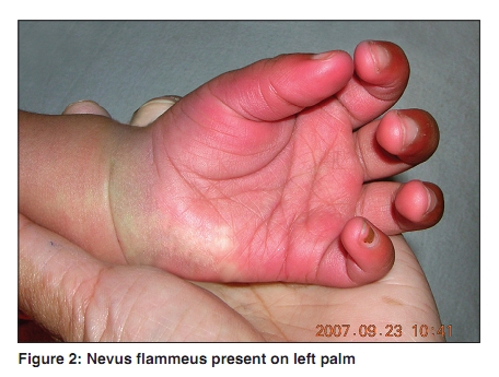
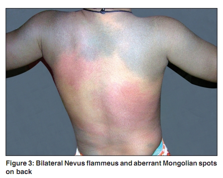
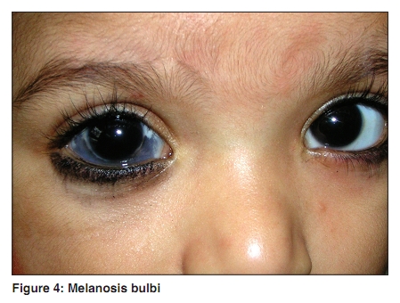
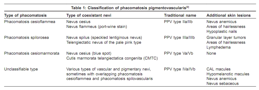
Indian Journal of Dermatology, Venereology, and Leprology, Vol. 76, No. 3, May-June, 2010, pp. 307 Net Case Phacomatosis cesioflammea: First case report from India Tarang Goyal, Anupam Varshney1 Departments of Dermatology and STD and 1Pathology, Muzaffarnagar Medical College, Muzaffarnagar, Uttar Pradesh, India Correspondence Address: Dr. Tarang Goyal, 105 Somdutt Vihar, Meerut, Uttar Pradesh - 250 004, India, tarang_derma@yahoo.co.in Code Number: dv10098 PMID: 20445318 DOI: 10.4103/0378-6323.62973 Abstract Phacomatosis cesioflammea is a rare condition defined by the simultaneous presence of both vascular and pigmentary nevus in the same patient. We report a case of a 4-year-old Indian female child who presented with diffuse dermal melanosis on the upper shoulders, upper anterior chest and lower back and extending to involve both sides of the arms and forearms, generalized port-wine stain on the back, shoulders and both upper limbs with sparing of the right palm. At places, the two types of lesions were superimposed on each other and were also present discretely on the back, but in close proximity to each other. There was the presence of melanosis bulbi on the right side of the eye. She was otherwise normal. She was clinically diagnosed as a case of phacomatosis pigmentovascularis cesioflammea. The nonallelic twin spotting phenomenon has been proposed in the pathogenesis of this disorder.Keywords: Phacomatosis cesioflammea, phacomatosis pigmentovascularis, melanosis bulbi, phenomenon, twin spotting Introduction Phacomatosis pigmentovascularis (PPV) is a very rare disorder with only about 200 cases being reported worldwide, mostly from Japan. [1] The most common type seen is phacomatosis cesioflammea. It was first described by Ota et al. in 1947 in detail as a rare combination of cutaneous hemangioma and pigmentary nevus and explained on the basis of the twin spotting phenomenon. PPV was further reclassified by Happle [2] into four subtypes in 2005. Phacomatosis cesioflammea has still not been reported from India. We report the case as being the first to be reported from India. Case Report A 4-year-old Indian female child presented with the following skin lesions in our department, which were noticed at or soon after birth: (1) numerous bilateral widespread reddish-pink macular lesions (nevus flammeus) over the left side of the upper anterior chest and upper back, continuing to involve both the arms, forearms and the left hand. [Figure - 1] and [Figure - 2], (2) additionally, she had aberrant large, diffuse, well-defined blue-black asymptomatic macular dermal pigmentary lesions (nevus fuscoceruleus) superimposed on these lesions with sparing of the right palm [Figure - 3]. These were also present on the lower back. On the back, both nevus flammeus and nevus fuscoceruleus were present independently, but in close proximity to each other. All the lesions were present since birth and were stationary in nature, (3) the sclera of the right eye showed bluish-black pigmentation - melanosis bulbi [Figure - 4]. The patient had no other complaint apart from cosmetic ones. On inspection, the hair, nail and mucus membranes were normal; no significant family history was elicited. All the limbs were symmetrical and no edema was seen. On tonometric examination, intraocular pressure was normal. Slit lamp examination was also normal. Family history is noncontributory. There was no history of consanguinity in her parents. Neurologic examination was normal. Regular follow-up of such patients should be carried out for ocular involvement or asymmetrical limb length development in later life. Discussion The Greek word "phacos" means "nevus." The term "phacomatosis" was previously mainly applied to genetically determined diseases of tissues of ectodermal origin, involving the central nervous system, eyes and skin. Now this term, together with a specifying adjective, is applied to few genetically determined diseases characterised by multiple nevi being present with or without systemic involvement, e.g., neurofibromatosis, tuberous sclerosis and PPV. PPV is defined as an association of a widespread vascular nevus with an extensive pigmentary nevus. Types described thus far can be explained on the basis of the twin spotting phenomenon. First case of the disorder was described in the 1920s and it was in 1947 that Ota et al. defined in detail a rare combination of cutaneous hemangioma and pigmentary nevus as PPV. The disorder was first subclassified into two types: type I, nevus flammeus and nevus pigmentosus et verrucosus and type II, nevus flammeus with aberrant Mongolian spots. In 1966, Toda described coexisting nevus flammeus and nevus spilus as type III. The fourth type was described by Hasegawa and Yasuhara in 1979 as a combination of nevus flammeus, aberrant Mongolian spots, nevus spilus and nevus anemicus. [1] In 2005, Rudolph Happle [2] reclassified it into three types: phacomatosis cesioflammea (identical with traditional types IIa and IIb), phacomatosis spilorosea (traditional types IIIa and IIIb) and phacomatosis cesiomarmorata (traditional type V) [Table - 1]. He further dropped the term type I and proposed to put the extremely rare unclassifiable form as type IV. Further, he proposed the term "phacomatosis multiplex" for some cases that could not be ascribed to any well-defined clinico-genetic entity. For example, Joshi et al.[3] described a combination of port-wine stain, congenital Becker's nevus, cafι-au-lait (CAL) macules and lentigines as a case of PPV type Ia, which can now be ascribed to be phacomatosis multiplex. Similarly, Chen and Happle [4] described a case that had a combination of telangiectatic nevi, blue nevi, CAL macules, nevus depigmentosus and nevus sebaceous in the same patient. In our case, we did not come across any other such abnormality as encountered by the above authors. Phacomatosis cesioflammea is the most common type of all PPVs. Vidaurri de la Cruz and colleagues [5] did not find any other type in a series of 24 consecutive cases. Associated systemic abnormalities that may be noticed are central nervous system defects, melanosis bulbi, glaucoma, asymmetrical length of limbs, dysplastic veins or lymph vessels and nevus anemicus. In addition, Sturge-Weber and Klippel-Trenaunay syndromes can be associated with this sub-type. The Latin word "caesius": blue-grey, serves as equivalent to the term "fuscoceruleus" to describe an aberrant Mongolian spot. It results from the entrapment of melanocytes in the dermis during their migration from the neural crest into the epidermis. In our case, the two types of nevi are in close proximity to each other, supporting the nonallelic inheritance of twin spot phenomenon. As both nevus flammeus and Mongolian spot arise from different cell types, it is assumed that autosomal recessive mutations are present on two different neighbouring loci and that they are exchanged simultaneously by somatic recombination. Twin spots consist of two genetically different clones of neighbouring cells in a background of normal cells. Two types of patterns are recognised: allelic (consisting of afferent cells of same stem cell lineage) and nonallelic (afferent cells of different stem cell lineage on a background of normal cells), and are represented by two paired areas with different phenotypes. [6] Examples of the nonallelic variant are PPV, phacomatosis pigmentokeratotica, Proteus syndrome and cutis tricolor. Apart from associated melanosis bulbi, no other associated anomaly was found. The presented case is unique as it is the first case reported from the Indian subcontinent. [7] References
Copyright 2010 - Indian Journal of Dermatology, Venereology, and Leprology The following images related to this document are available:Photo images[dv10098f4.jpg] [dv10098f3.jpg] [dv10098f1.jpg] [dv10098f2.jpg] [dv10098t1.jpg] |
| |||||||||
