 |
| About Bioline | All Journals | Testimonials | Membership | News |
|
||||||
|
||||||
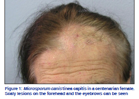
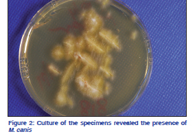
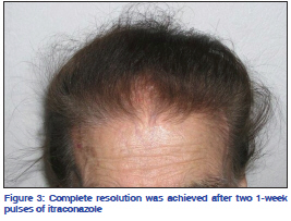
Indian Journal of Dermatology, Venereology, and Leprology, Vol. 77, No. 5, September-October, 2011, pp. 626 Net Letter Microsporum canis tinea capitis in a centenarian patient Efstathios Rallis, Elma Koumantaki-Mathioudaki1, Helen Papadogeorgakis2 Department of Dermatology, Veterans Administration Hospital (NIMTS), Athens, Departments of 1Dermatology and 2Mycology, University of Athens, “A. Sygros” Hospital, Athens, Greece Correspondence Address: Code Number: dv11190 DOI: 10.4103/0378-6323.84077 Sir, Tinea capitis is a dermatophyte infection of the scalp and hair. It is overwhelmingly an infection of prepubertal children, but is being increasingly recognized in adults though it was once considered to be uncommon in this age-group.[1] Microsporum canis (M. canis) is the dominant cause of tinea capitis in Greece.[2] In this article we present probably the oldest patient with M. canis tinea capitis to be reported in the literature. A 100-year-old immunocompetent female was referred to our hospital because of scaly alopecia with pustules and crusts of 2 months duration [Figure 1]. Hairs in the affected area were gray and breaking off just above the level of the scalp. Scaly lesions on the forehead and the eyebrows, resembling seborrheic dermatitis, could also be seen. The lesions had not responded to oral amoxicillin and topical fusidic acid, which had been prescribed by her physician. The patient had a body weight of 48 kg. She lived in a village in the mountains. She mentioned no contact with animals and did not use any clothing for the head. No fungal infections were detected on other parts of her skin or her nails and hair and none of her close family members had been similarly affected. Despite her age, the medical history was unremarkable apart from a history of chronic gastritis for which she was not receiving any medication. She had no evidence of any sort of immunological deficiency. Culture of bacterial swabs was negative. Mycological examination was performed on the scales from the lesion and hair samples. Direct examination of a 20% KOH preparation was positive. The specimens were also inoculated on Sabouraud 2% glucose– chloramphenicol agar and Sabouraud 2% glucose cycloheximide–chloramphenicol agar and revealed the presence of the ectothrix M. canis [Figure 2]. The scalp of the patient was also examined under a Wood’s lamp and it fluoresced a green color. We decided to treat her with itraconazole pulse therapy. Each pulse comprised of capsule itraconazole 100 mg (Verisfield, Athens, Greece) twice daily for 1 week, with an interval of 3 weeks between the pulses.[3] To avoid gastrointestinal symptoms and/or exacerbation of gastritis due to itraconazole and because of her low body weight, we decided to administer a dose of 100 mg of the medication for three pulses instead of the standard 5 mg/kg/day; in case of treatment failure, we would switch to a continuous regimen of 100 mg itraconazole. After 2 months of pulse therapy she called us over the phone as she was not able to visit our department. She reported that her hair had gradually grown back again and we suggested that she discontinue the treatment. Three months after discontinuation of the treatment she visited the department again. There was complete remission of the dermatophyte infection; all scaly lesions had subsided and there was no evidence of recurrence [Figure 3]. Mycological examination was negative. She did not report any adverse events during the treatment period. Dermatophytic colonization of the scalp usually disappears at puberty. Tinea capitis in adults has been reported to occur in patients who are immunosuppressed or HIV-infected;[4] however, there is not enough evidence to support this. Devliotou- Panagliotidou et al.[1] reported 35 adults with tinea capitis, 27 of them with a typical pattern of menopause but none with any immunological deficiency. A female predominance in the adult cases has been reported but has not been explained.[5] Numerous factors have been incriminated, such as contact with affected family members, hormonal differences, the composition of sebum, and immunological deficiency. Griseofulvin has been the gold standard for the treatment of tinea capitis. Little data is available about the efficacy of fluconazole in tinea capitis.[6] Terbinafine has been licensed since 1996 for the treatment of mycotic infections of the scalp in children and adults.[7] However, the cure rates with terbinafine are poorer when the causative organism is M. canis than when it is T. tonsurans and T. violaceum.[8] Itraconazole is probably the ideal alternative to griseofulvin for fungal infections of the scalp.[3] Our patient responded very well to the oral pulse therapy regimen of itraconazole. Itraconazole represents a good therapeutic choice for ectothrix fungal infections of the scalp. In such cases, concomitant use of ketoconazole shampoo 2–3 times a week is also recommended to prevent fomite transmission. REFERENCES
Copyright 2011 - Indian Journal of Dermatology, Venereology, and Leprology The following images related to this document are available:Photo images[dv11190f3.jpg] [dv11190f2.jpg] [dv11190f1.jpg] |
| |||||||||
