 |
| About Bioline | All Journals | Testimonials | Membership | News |
|
||||||
|
||||||
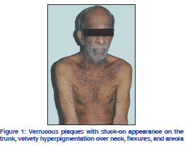
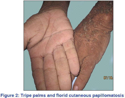
Indian Journal of Dermatology, Venereology, and Leprology, Vol. 77, No. 5, September-October, 2011, pp. 626 Net Letter A myriad of paraneoplastic dermatoses Smitha A. Varghese, Sobhanakumari K., Celin M. Issac, Seena P. Department of Dermatology, Medical College, Kottayam, Kerala, India Correspondence Address: Code Number: dv11193 DOI: 10.4103/0378-6323.84081 Sir, Paraneoplastic dermatoses, describes those benign disorders in which there is a direct and often parallel course of the dermatosis with an underlying malignancy. Their presence serves as an important marker of a potentially-associated neoplastic process. We are reporting a case with multiple cutaneous paraneoplastic manifestations seen in association with an upper gastrointestinal (GI) malignancy. A 76-year-old man presented with generalized, pruritic, hyperpigmented raised lesions for 4 years and hyperpigmentation over the sides of neck, axilla, and thighs for 4 years. On examination, patient appeared emaciated; pallor was present and there was no significant lymphadenopathy or systemic findings. He had hyperpigmented velvety plaques over neck, axilla, groin, areola, and lips suggestive of acanthosis nigricans (AN); numerous verrucous papules and plaques with a stuck-on appearance over trunk, face, scalp, and proximal extremities suggestive of seborrheic keratosis [Figure 1]; palms showed hyperkeratosis and altered dermatoglyphics suggestive of tripe palms and verrucous papules typifying verruca vulgaris seen over extensor surfaces of extremities [Figure 2]. On detailed enquiry, patient revealed that he was a chronic smoker for the past 40 years. He also had difficulty in swallowing solid foods since 6 months, and had loss of weight and appetite since 3 months. On investigation, hemogram showed an hemoglobin of 7.8 g/dl with raised erythrocyte sedimentation rate, urine routine examination was normal and stool occult blood was negative. Liver function tests, renal function tests, peripheral smear, and chest X-ray were within normal limits. Biopsies were taken from the palms, stuck-on lesions of back and verrucous plaques and were consistent with the diagnosis of tripe palms, seborrheic keratosis and florid cutaneous papillomatosis, respectively. As part of the work up for internal neoplasia, Ultrasound abdomen showed minimal wall thickening in the cardio-esophageal junction with an upper para-aortic node. Computerized tomography of abdomen and thorax showed a growth at the distal esophagus extending to the proximal stomach and enlarged para-aortic nodes. Trachea, bronchi, and lung parenchyma were normal. Oesophago-gastroduodenoscopy showed a growth at the lower end of esophagus obstructing further passage of scope into stomach. Biopsy from the growth revealed a squamous cell carcinoma of the lower end of esophagus of poorly differentiated type. Owing to the advanced stage of the disease, our patient could only be offered palliative treatment. The four paraneoplastic dermatoses described in our patient, ie, acanthosis nigricans, tripe palms, leser trelat sign, and florid cutaneous papillomatosis belong to a common spectrum of proliferative paraneoplastic disorders and should be viewed as part of a continuum. These conditions develop by means of a common pathogenic pathway due to an underlying malignancy that produces a factor similar to human epidermal growth factor.[1] One hypothesis suggests that the secretion of large amounts of transforming growth factor α(TGF-α) by the tumor into the circulation stimulates keratinocyte growth via an endocrine route. TGF-α is closely related to epidermal growth factor (EGF) and binds to the same receptor, EGFR. This binding induces activation of the classical mitogenactivated protein kinase (MAPK, ERK) pathway, a module of three protein kinases that is organized in a hierarchical fashion and is known to regulate basic cellular functions such as proliferation, differentiation, and migration.[2] Another hypothesis states that germline mutations in fibroblast growth factor, FGFR3 also induce a proliferative effect on keratinocytes. Strong expression of FGFR3 was frequently observed in seborrhoeic keratoses and acanthosis nigricans by immunohistochemical analysis, and that activation of FGFR3 might be a common feature in tumorigenesis in seborrhoeic keratoses and malignant acanthosis nigricans. FGFR3 might also contribute to ras/MAPK signaling in the lesions, because EGFR and FGFR3 are both transmembrane tyrosine kinase receptors (TKRs) and located upstream of the RAS/MAPK pathway.[3] Further investigations are needed to elucidate how EGFR and FGFR3 cooperate in the pathogenesis of these cutaneous proliferative disorders. There have been reports suggesting coexistence of acanthosis nigricans and the sign of Leser-Trélat and florid cutaneous papillomatosis in patients with gastric adenocarcinoma[4,5] which is the commonly associated malignancy but in our case the associated primary is a squamous cell carcinoma of the esophagus which is unusual. REFERENCES
Copyright 2011 - Indian Journal of Dermatology, Venereology, and Leprology The following images related to this document are available:Photo images[dv11193f2.jpg] [dv11193f1.jpg] |
| |||||||||
