 |
| About Bioline | All Journals | Testimonials | Membership | News |
|
||||||
|
||||||
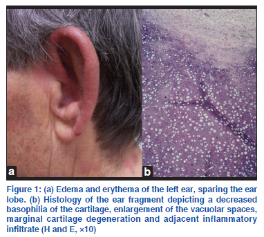
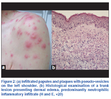
Net Letter Paraneoplastic relapsing polychondritis and Sweet syndrome coexisting in a patient with myelodysplasia Ana M. Calistru1, Carmen Lisboa1,2, Filomena Azevedo1 1Department of Dermatology and Venereology, Hospital São João,
EPE, Porto, 2Faculty of Medicine (U38/FCT),
University of Porto, Portugal Code Number: dv11225 Sir, Relapsing polychondritis (RP) is a rare inflammatory systemic disorder with cartilaginous tissues, such as ears, nose, joints and respiratory tract, as primary targets. Association of RP with hematological malignancies, particularly with myelodysplastic syndrome (MDS), has been described.[1] Sweet syndrome (SS) is an acute febrile neutrophilic dermatosis that may be related with neoplasm, especially hematological. A 71-year-old man was referred to us for pain, edema and erythema of the left ear, sparing the ear lobe [Figure 1a] and hand arthralgias, evolving for 3 weeks. He has been treated unsuccessfully with antibiotics for suspected infectious perichondritis. Two days prior to admission, the patient became febrile (38°C), developing erythematous infiltrated papules and plaques with pseudo-vesicles on his face, trunk and upper limbs [Figure 2a]. There was no hearing loss, nor ocular or respiratory complaints. The patient was diagnosed with MDS 3 years before and was treated with blood transfusions and desferrioxamine. A biopsy of the ear lesion displayed a decreased basophilia of the cartilage, enlarged vacuolar spaces, marginal cartilage degeneration and adjacent inflammatory infiltrate [Figure 1b], consistent with chondritis. The trunk lesion histology revealed dermal edema, predominantly neutrophilic inflammatory infiltrate and negative direct immunofluorescence, compatible with SS [Figure 2b]. The laboratory workup found pancytopenia, elevated C-reactive protein (42 mg/l; normal <3), a 1/100 titer of antinuclear antibodies (ANA) (normal<1/100) with homogeneous pattern, and an elevated titer of Anti-double stranded Deoxyribonucleic acid (anti-dsDNA) antibodies (31.7 UI/ml; normal<20) detected by Enzyme-linked immunosorbent assay (ELISA). The rheumatoid factor and the anti-extractable nuclear antigen antibodies were negative. The microbiologic examination of the ear fragment ruled out bacteriological, mycological and mycobacteriological infections. The head and neck computed tomography (CT)-scan showed no upper respiratory tract compromise. In addition, the immunophenotypic analysis of the peripheral blood and bone marrow examination did not indicate leukemia progression. Evidence of auricular chondritis with compatible histology features and inflammatory polyarthritis led to the diagnosis of RP, according to the Damiani and Levine criteria.[2] Concomitant SS diagnosis was achieved with two major criteria (sudden onset of specific lesions and consistent histopathology) and two minor criteria (associated hematological disorder and fever). The patient received Ibuprofen (400 mg, three times daily orally) and MDS treatment was optimized with the introduction of erythropoietin. Improvement of the RP symptoms was achieved after 2 weeks. The SS lesions resolved with potent topical steroid therapy (betamethasone dipropionate 0.05% cream, twice a day for 10 days). After a follow-up of 10 months, there was no record of relapse or new symptoms and the laboratory workup showed persistence of elevated anti-dsDNA (57 UI/ml) and borderline ANA antibodies (1/100). The etiology of RP is unknown, although there is evidence for autoimmune pathogenesis: the presence of T-cells infiltrate and antigen-antibody complexes in affected cartilages and circulating antibodies to cartilage-specific collagen types II, IX, XI. RP may occur in a primary form or in association with other disorders, such as autoimmune diseases and malignancies, where it is regarded as paraneoplastic.Considering the present report, the association of RP and SS is documented in 23 cases.[1,3-6] Fourteen of these patients had a related malignancy: twelve had MDS, one had chronic lymphocytic leukaemia and another one had bladder cancer. Thus, it is conceivable that the development of SS and RP in the same patient is more than a mere coincidence,[1] implying a common cause or pathogenesis possibly related to neutrophil infiltration.[6] Immunological imbalances due to MDS concurrent with a specific immunogenetic background may play key roles in the pathogenesis of RP in these patients. Autoimmune paraneoplastic syndromes or asymptomatic immunologic abnormalities may be encountered in patients with MDS. Our patient depicted an elevation of anti-dsDNA antibodies without fulfilling the criteria for systemic lupus erythematosus. The anti-dsDNA antibodies detected by ELISA may be present in up to 30% of other disease groups, such as rheumatoid arthritis, or in normal subjects. Accordingly, the elevated anti-dsDNA and the borderline ANA antibodies in our patient may represent either autoimmune manifestations related to RP or immunologic abnormalities associated to MDS. Systemic corticosteroids and non-steroidal antiinflammatory drugs are the mainstay of RP therapy.[1] We decided to avoid systemic corticosteroid therapy due to the lack of multi-organ involvement and due to the additional risk of immunosuppression. Resolution of the symptoms was achieved through oral nonsteroidal anti-inflammatory treatment for chondritis and arthralgias, and with potent topical steroid for SS lesions. Herein, we report the association of two paraneoplastic syndromes with cutaneous expression in a patient with myelodysplasia. Our case also demonstrates the importance of recognizing RP in the differential diagnosis of an infectious perichondritis. Further knowledge of the pathogenesis of RP, SS and MDS will provide important insights regarding the coexistence of these diseases. REFERENCES
The following images related to this document are available:Photo images[dv11225f1.jpg] [dv11225f2.jpg] |
| |||||||||
