 |
| About Bioline | All Journals | Testimonials | Membership | News |
|
||||||
|
||||||
Electronic Journal of Biotechnology, Vol. 5, No. 3, December, 2002 Choice of the adequate quantification method for recombinant human GM-CSF produced in different host systems Mariela Bollati Fogolín1, Marcos Oggero Eberhardt2, Ricardo Kratje3, Marina Etcheverrigaray* 4 1Laboratorio
de Cultivos Celulares, Facultad de Bioquímica y Ciencias Biológicas,
Universidad Nacional del Litoral, Ciudad Universitaria - C.C. 242, S3000ZAA Santa
Fe, Argentina, Tel: 54 342 4575 214, Fax: 54 342 4575 214, E-mail: mrb@gbf.de Finanacial support: This work was supported by a grant from the Agencia Nacional de Promoción Científica y Tecnológica (Argentina), BID 802, PID Nr. PMT-SID 187. Received
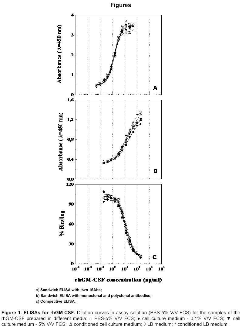
September 13, 2002 / Code Number: ej02038 Abstract Three enzyme-linked-immunosorbent assays (ELISA) were developed and compared with a bioassay to quantify the recombinant human granulocyte-macrophage colony stimulating factor (rhGM-CSF). These assays were suitable to quantify the non-glycosylated rhGM-CSF present in mixtures with variable protein content, and therefore useful for determining concentrations of the cytokine in production processes. Among these assays, the competitive ELISA, developed with a MAb that recognises an epitope not involved in glycosylation, was the only one suitable to quantify the glycosylated form of rhGM-CSF produced in mammalian cell cultures. Keywords: bioassay, ELISA, GM-CSF, monoclonal antibody, quantification. Abbreviations: rhGM-CSF: recombinant human granulocyte-macrophage colony stimulating factor; ELISA: enzyme-linked-immunosorbent assays; MAb: monoclonal antibody; RIA: radioimmnunoassay; PAb: polyclonal antibody; FCS: foetal calf serum; PBS: phosphate buffered saline; LB: Luria-Bertani medium; HRP: horseradish peroxidase; OPD: o-phenylenediamine; CV: coefficients of variation; CHO: Chinese hamster ovary; SEM: standard error of the mean. Article The human granulocyte - macrophage colony stimulating factor (hGM-CSF) is a hematopoietic growth factor which mediates the differentiation and proliferation of granulocyte and macrophage colonies in the bone marrow (Metcalf, 1985). There is a lot of clinical interest in this lymphokine due to both its ability to stimulate the granulocyte and macrophage production in patients who are immunosuppressed -either from disease or from receiving chemotherapy or radiation therapy- and also its potential in treating myeloid leukemia. The hGM-CSF has been introduced into clinical treatment with promising results (Hussein et al. 1995). The hGM-CSF in its natural form is a variable glycosylated protein with a reported mol wt of 15,000 to 31,000 daltons (Nicola, 1994). Following the cloning of the human GM-CSF gene, the biosynthetic hGM-CSF has been produced and purified from yeast, mammalian cells and bacteria, resulting in proteins that vary in composition, glycosidic content and structure (Dorr, 1993). During the production process of this hematopoietic growth factor, specific quantitative assays are required. Factor-dependent cell lines have been established and used in measurements of the hGM-CSF biological activity. A RIA has also been developed for detecting fluctuation of the GM-CSF, both in health and in disease (Thing Mortensen et al. 1993), and an ELISA has been described to determine the pharmacokinetics of the bacterially synthesised hGM-CSF in phase I studies (Cebon et al. 1988). Here, we report the development and comparison of different assays carried out in order to quantify the cytokine during a production process. Three ELISAs for the rhGM-CSF were developed to quantify the rhGM-CSF derived either from E. coli or mammalian cells and compared with the bioassay based on the proliferation of TF-1 cells. Materials and Methods GM-CSF preparations. The glycosylated rhGM-CSF was expressed in COS and CHO.K1 cells using a plasmid which includes the Adenovirus major late promoter, by lipofection (Etcheverrigaray et al. 1998; Bollati Fogolín et al. 2000). The E. coli - derived hGM-CSF purchased from Schering-Plough Co. (Ireland) was the non-glycosylated rhGM-CSF form and it was used as an internal standard, previously assayed against the E. coli - derived hGM-CSF International Standard (NIBSC, 88/646, UK). Antibodies. Hybridoma clones secreting monoclonal antibodies (MAbs) against the rhGM-CSF were generated in BALB/c mice according to standard protocols (Galfrè and Milstein, 1981). Ascitic fluid was obtained in mice. MAbs were evaluated for their ability to inhibit the proliferation of hGM-CSF stimulated TF-1 cells, i.e. to neutralise the in vitro hGM-CSF activity. MAb M1B8 and M7E10 were selected for ELISA tests taking into account their capacity to neutralise both glycosylated and non-glycosylated forms of the cytokine in the in vitro biological assay, their isotype (IgG1), as well as their capacity to recognise different epitopes (Oggero et al. 2001). Briefly, MAb M1B8 recognises the conformational epitope constituted by the aminoacids P118F119W13E14 and MAb M7E10, the peptide L61YKQGLRGSLTK72 that is part of a conformational epitope. GM-CSF polyclonal serum was obtained using a standard hyper immunisation protocol. Briefly, 50 µg rhGM-CSF in saline was emulsified with an equal volume of Freund’s complete adjuvant and injected subcutaneously into multiple dorsal sites in rabbits. The treatment was repeated 4 weeks later and the animals were bled 10 days after the last injection. In all cases, pure rhGM-CSF kindly provided by the company Genargen S.R.L. (Buenos Aires, Argentina) was used as immunogen. Monoclonal antibodies were purified from ascitic fluid with Protein A - Sepharose 4 FF (Amersham Biosciencies, Sweden) using the dealer´s guidelines. Biotinylations were performed as described by Bayer and Wilchek, 1980. Analysis of hGM-CSF in selected protein-rich samples. Samples consisted of non-glycosylated E. coli - derived rhGM-CSF diluted in different media, in order to evaluate the influence of the cytokine’s environment in its quantification: phosphate buffered saline containing 5% V/V foetal calf serum (PBS-5% FCS), mammalian cell culture medium containing 0.1 or 5% V/V FCS, cell culture supernatant (conditioned medium), Luria-Bertani medium (LB) and conditioned LB. In order to establish whether total protein concentration was critical regarding interference, dilution curves were performed in these media as well as in the dilution buffer used for the standard curve. Detection of the rhGM-CSF. The following quantification assays were developed: a sandwich ELISA with two MAbs, a sandwich ELISA with monoclonal and polyclonal antibodies, a competitive ELISA and a cell proliferation assay. All ELISA had the following common steps:
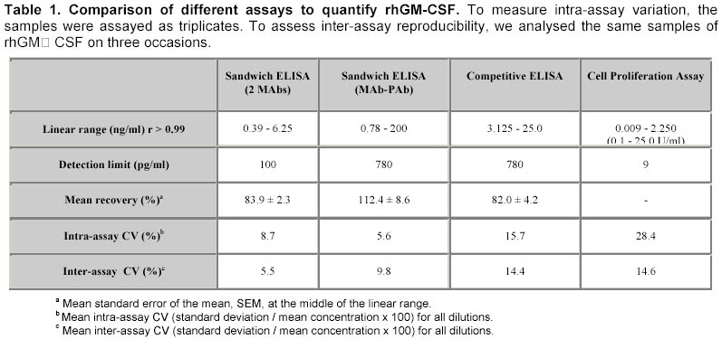
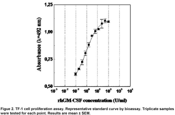
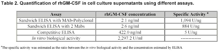
Sandwich ELISA with monoclonal and polyclonal antibodies. Each well was coated with 100 µl M1B8 ascitic fluid diluted 1:2,000. After washing, 100 µl standard rhGM-CSF in PBS-5% V/V FCS, over the range 0.195-200 ng/ml, or 100 µl samples in 1:2 serial dilutions were added to the wells. All incubations were carried out at 37ºC for 1h. Afterwards, plates were washed and 100 µl rabbit polyclonal antiserum diluted 1:1,000 in PBS-FCS-T was added and after incubation and washing, 100 µl of goat anti-rabbit immunoglobulins-HRP conjugate diluted 1:1,000 in the same buffer was added. Once more, plates were incubated and washed, and 100 µl of substrate was added. Optical densities were measured as described. Sandwich ELISA with two monoclonal antibodies. Wells were coated with 100 µl M1B8 ascites diluted 1:500. Plates were washed and 100 µl standard rhGM-CSF in PBS-5% V/V FCS over the range 0.049-50 ng/ml or 100 µl samples in 1:2 serial dilutions were added to the wells. Plates were washed and 100 µl biotinylated MAb M7E10 diluted 1:500 in PBS-FCS-T was added. After incubation, plates were washed and 100 µl streptavidin-HRP conjugate diluted 1:250 was added. Finally, 100 µl substrate was added and allowed to react. Optical densities were measured as described. Competitive ELISA. Wells were coated with 16.5 ng rhGM-CSF. It was also used as competitor in solution, serially diluted from 200 to 0.195 ng/ml in PBS-5% V/V FCS. Samples were assayed in serial 1:2 dilutions. Competition was carried out incubating the coated plates with 100 µl competitor or samples with 100 µl M7E10 ascites 1:100,000 in PBS-5% V/V FCS, at 37ºC for 2 h. After washing, 100 µl rabbit anti-mouse immunoglobulins-HRP conjugate 1:1,000 was added to the wells. After incubation for 1h at 37ºC, plates were washed and 100 µl substrate was added. Optical densities were measured as described. Cell proliferation assay. To quantify the biological activity of the rhGM-CSF, the factor dependent cell line TF-1 (ATCC CRL-2003) was used. TF-1 cell proliferation was determined measuring the activity of dehydrogenase-enzyme as marker for the biological activity, using 2 mg/ml Cell Titer 96TM MTS Reagent Powder (3-(4,5-dimethylthiazol-2-yl) -5-(3-carboxymethoxyphenyl) -2-(4-sulfophenyl) -2H-tetrazolium inner salt) (Promega, USA) and 0.92 mg/ml Phenazine methosulfate (Sigma, USA). TF1 cells were cultured under 5% CO2 in RPMI 1640 (Gibco BRL, USA) containing 2 mM L-glutamine, 0.05 mM ß-mercaptoethanol, 10% V/V FCS and 50 U/ml rhGM-CSF. For the assay, exponentially growing cells were collected and washed with the same culture medium lacking FCS and rhGM-CSF in order to eliminate the cytokine. 50 µl serial 1:2 dilutions of standard rhGM-CSF (in TF-1 cell culture medium lacking GM-CSF) was added to 96 well culture plates (NuncTM Brand Products, Denmark) from 100 - 0.78 U/ml and 50 µl of cell suspension containing 3.105 cells/ml was inoculated in each well. After incubation for 2 days at 37ºC under 5% CO2, 20 µl developing reagent was added and the incubation was continued for 3 h. hGM-CSF dependent cell proliferation was determined as the absorbance at 492 nm. Negative controls were performed with TF-1 cells cultured without rhGM-CSF. Results Dilution curves for detecting the rhGM-CSF in the different samples by sandwich ELISA with two MAbs are presented in Figure 1a. The recovery was variable, depending on the protein content of the samples as they were assayed diluted in the original medium (data not shown). In contrast, only small differences were detected between the standard curve and the dilution curves of the samples, developed in PBS-5% V/V FCS. Intra and inter assay coefficients of variation (CV = standard deviation / mean concentration x 100) were tested at concentrations of 5.0, 2.5 and 1.25 ng rhGM-CSF/ml. Table 1 summarises the results of all the methods used to quantify the non glycosylated rhGM-CSF. The results obtained by sandwich ELISA with polyclonal and monoclonal antibodies are shown in Figure 1b. The recovery percentage was variable -depending on the nature of the sample-, thus showing that the lower the protein environment of the sample, the lower the recovery (data not shown). Intra and inter-assay CV were tested at concentrations of 20, 10 and 5 ng rhGM-CSF/ml. Figure 1c shows the results of the competitive ELISA. No differences were detected between the standard curve and the dilution curves developed in different media (data not shown). The protein environment of the sample does not modify the values determined by ELISA. Intra and inter-assay CV were tested at concentrations of 20, 10 and 5 ng rhGM-CSF/ml. The results of the cell proliferation assay are shown in Figure 2. Intra and inter-assay CV were tested at concentrations of 140, 70 and 35 pg rhGM-CSF/ml. When the three ELISA were used to quantify the glycosylated rhGM-CSF present in mammalian cell culture supernatants, they provided different concentrations of the cytokine and consequently, different values of specific activity were estimated (Table 2). Only the competitive ELISA provided a value of specific activity comparable with those previously reported (Cebon et al. 1988). Discussion We have described rapid and reliable immunoassays for rhGM-CSF in mixtures with variable protein content. These ELISA were developed with the aim of devising quick and sensitive assays without the use of radioisotopes and bioassays. Bioassays are laborious, lengthy and subjected to inherent inaccuracies. Cell line based bioassays are prone to problems that need addressing before reliable estimates of cytokine levels can be derived. The sample may contain dilutable non-cytokine components that affect the assay, a problem often encountered when using samples that contain serum components. Continuously growing cell lines can change their cytokine responsiveness if cultured for long periods. Mycoplasma contamination can also drastically reduce the response of cell line based bioassays. Different batches of foetal calf serum used to maintain cultures can also affect the performance of bioassays and should be carefully screened prior to use (Mire-Sluis and Thorpe, 1998). Therefore, alternative methods are needed for the rapid screening of a large number of samples. The assays were developed using non-glycosylated rhGM-CSF, and they were intended to be used in the production process by mammalian cell culture, which permits the obtainment of the glycosylated molecule. All the methods are sensitive enough for the detection of the non-glycosylated cytokine in the upstream and the downstream processes. Among the ELISAs developed in this work, the sandwich ELISA with two MAbs is the least influenced by the protein environment, taking into account the low deviation of the recovery when samples are diluted in assay solution. This method shows also the lowest CV and becomes the most appropriate assay to quantify non-glycosylated GM-CSF. The sandwich ELISA with monoclonal and polyclonal antibodies has the broadest linear range. Considering that these monoclonal antibodies also recognise the CHO-derived cytokine (they neutralise its in vitro biological activity, as previously described), an ELISA that involves the use of such antibodies may be able to quantify the glycosylated molecule. All antibodies used in the development of these assays recognise both the non-glycosylated and the glycosylated cytokine. However, experiments carried out with the aim of quantifying by ELISA the glycosylated rhGM-CSF in cell culture supernatants after the transfection of mammalian cells, showed that different results are obtained by the three described ELISAs. In this way, the competitive ELISA is the only assay useful to quantify glycosylated rhGM-CSF. A previous work demonstrated that MAb M7E10 used in this assay has similar affinity for both, glycosylated and non-glycosylated rhGM-CSF (Oggero et al. 2001), mapping a region not involved in the glycosylation. Considering previous data from the literature, which describe specific activities between 5-12 UI/ng for both forms of this molecule (Mire-Sluis et al. 1995b), it can be argued that the competitive ELISA which uses only one particular MAb is more accurate in determining the glycosylated molecule. It confirms the importance of the characterisation and selection of antibodies to be used in the development of immunochemical methods. It is possible that the need of only one epitope for the reaction plays a role in this result. Sandwich assays need at least two different epitopes to be recognised by the monoclonal or polyclonal antibodies. It may be limiting in the case that the epitopes are partially masked by glycosidic residues, as in the case of glycosylated cytokines. It may be explained as a steric hindrance due to the high glycosidic content of this protein. Moreover, when the protein is immobilised on the solid surface after reaction with the immobilised MAb, the conformational change that the antigen suffers can affect the reaction with the second antibody. The measurement of the biological activity of this cytokine may be done in vitro by proliferation assays that use hGM-CSF responsive cell lines. The nature of the reference standard in a bioassay is of considerable importance. Standards calibrated in mass are of little value since the specific activity of different recombinant preparations of cytokines differ and are most likely to be different from the specific activity of naturally occurring materials (Mire-Sluis et al. 1995a and Mire-Sluis et al. 1995b). In this case, there is no international standard for the CHO-derived rhGM-CSF and therefore, it cannot be quantified by bioassay. Therefore, the developed competitive ELISA becomes the method of choice for the quantification of the glycosylated form of this cytokine during its production process, from cell supernatants to the purified end product. References
Note: Electronic Journal of Biotechnology is not responsible if on-line references cited on manuscripts are not available any more after the date of publication. Supported by UNESCO / MIRCEN network © 2002 by Universidad Católica de Valparaíso -- Chile The following images related to this document are available:Photo images[ej02038f2.jpg] [ej02038t2.jpg] [ej02038t1.jpg] [ej02038f1.jpg] |
| |||||||||
