 |
| About Bioline | All Journals | Testimonials | Membership | News |
|
||||||
|
||||||
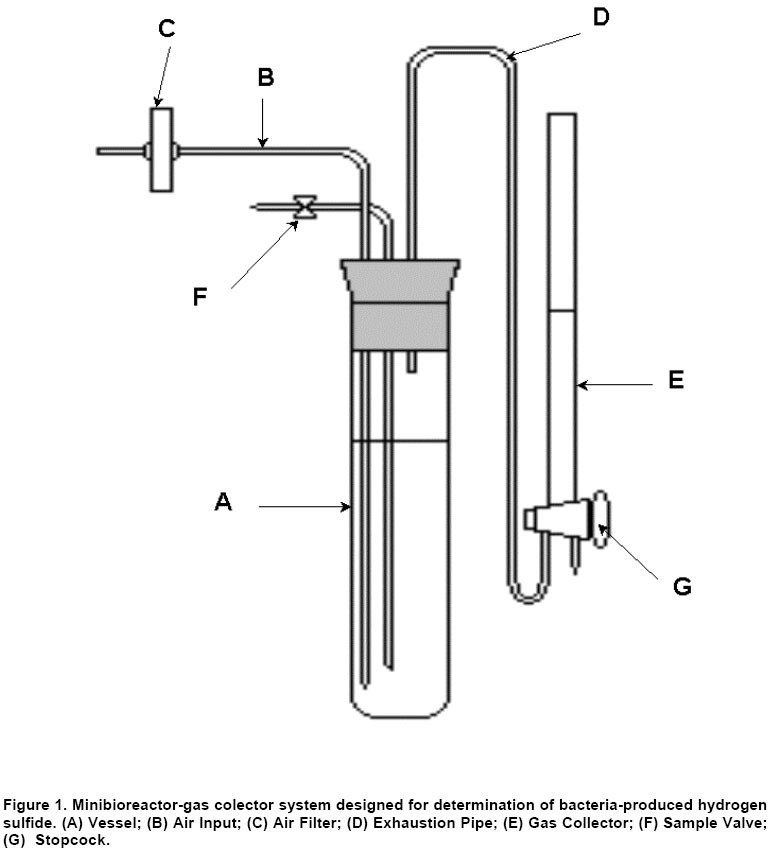
Electronic Journal of Biotechnology, Vol. 6 No. 3, December 15, 2003 Minibioreactor-gas collector for determining bacteria-produced hydrogen sulfide Armando Hernández García*1, Eulogio Pimentel Vázquez2, Jesús Mena Campos3 1Centro
de Ingeniería Genética y Biotecnología de Camagüey,
P.O. Box 387, Camagüey 70100, Cuba,
Tel: 5332261014/5332261024
Fax: 5332261587
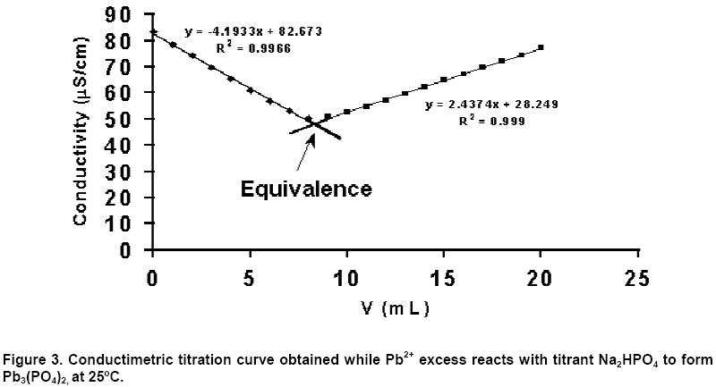
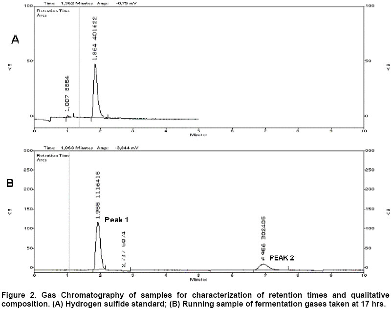
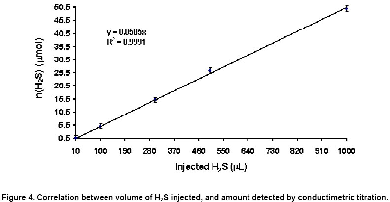
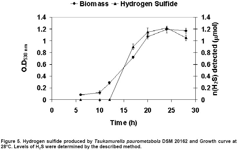
E-mail: armando.hernandez@cigbcam.cigb.edu.cu Financial support: This work was part of a project financed by Ministry of Sciences, Technologies and Environment of Cuba. Received January 20, 2003 / Code Number: ej03026 Abstract A simple and economical minibioreactor-gas collector system for determination of hydrogen sulfide produced by a microorganism was designed. The detection of hydrogen sulfide was based on the reaction between the hydrogen sulfide in the gas stream from the culture, and a lead acetate solution 0.090 mol/L, contained in a tube gas collector; the conductimetric back titration of lead excess was made, and hydrogen sulfide was quantified indirectly, being the detection limit 0.5 µmol. The developed system was applied using Tsukamurella paurometabola DSM 20162 as a model, being the amount of hydrogen sulfide produced, 1.2 µmol in 24 hrs. Keywords: bacteria, bacterial growth, conductimetric titration, microorganisms, sulfur-containing gases. Abbreviations: rpm: revolutions per minute. There is a great diversity of microorganisms that produce hydrogen sulfide (Aiking et al. 1982; Kim and Olson, 1989; Barton and Tomei, 1995; Cooney et al. 1996; Levine et al. 1998; Wang et al. 2000), from organic (i.e. sulfur-containing amino acids) and inorganic sources (i.e. sulfates). Although hydrogen sulfide is a toxic pollutant gas generally occurring in wastewater, it has been used to precipitate metals in wastewater treatment reactors and has been proposed for stabilization of metals in soils and for formation of metal sulfide "quantum" particles for microelectronics applications (Holmes et al. 1997). When a novel hydrogen sulfide producer bacterial strain is isolated, it is necessary the physiological and biochemical characterization of such strain, in order to optimize the hydrogen sulfide production for its further application (Fortin et al. 1994; Peyton et al. 1995; White and Gadd, 1996; White and Gadd, 1998; Smith, 2000). Although the experimental determination of this compound has been done through different methods, and the analytical methods for H2S detection and quantification have ranged from spectrophotometric analysis (Cline, 1969; Acree et al. 1971; Siegel, 1965; Chu et al. 1997) to gas chromatography (GC), either with distinct features in terms of accuracy, precision and detection limit (Heida et al. 1995; Chinivasagam et al. 1998; Mestres et al. 1999); the study at small scale is a suitable and economical option for the achievement of a such objective. In this paper a minibioreactor-gas collector system designed for characterization and study of hydrogen sulfide production from bacteria, is described. In addition, a simple analytical method, based on H2S indirect conductimetric determination as a modification of the method of lead acetate for detection of hydrogen sulfide (Hunter and Crecelius, 1938), was developed. The gas stream from the minibioreactor passes through a lead acetate solution contained in the tube (gas collector), thus H2S reacts with Pb2+, producing lead sulfide; then the Pb2+ excess is determined by conductimetric titration with sodium phosphate. Materials and Methods Development of the Analytical Method H2S standard preparation. In order to develop a method of analysis, a H2S standard was prepared, from the reaction of 1 g of Na2S (Merck) with 11 mL of 0.1 N H2SO4 (BDH), in a tightly closed 100 mL bottle. The bottle was sealed immediately after the Na2S was added, then H2SO4 was injected into the bottle. Method description. 10 µL, 100 µL, 300 µL, 500 µL, 700 µL and 1000 µL of the H2S previously prepared standard were injected into different sealed bottles containing 2 mL of 0.090 mol/L Pb(CH3COO)2 (Merck) solution, which produced a black PbS precipitate. The content in each bottle was filtered with Whatman paper 40, and transferred to 250 mL volumetric flasks and filled up. Each point had 5 replicas. Volumes of 100 mL were taken from the volumetric flasks and titrated with a Na2HPO4 solution (Merck) 4 mM; conductivity was measured for each volume of added titrant with a Copenhagen CDM 80 Radiometer Conductivity Meter. The conductimeter was calibrated using standard solutions of KCl (Radiometer) with conductivities 20 µS/cm and 100 µS/cm respectively at 25ºC. In each particular case, the equivalence point was determined from the conductimetric titration curve (Figure 3). Once the curves were adjusted by linear regression (descending and ascending straight lines) the equivalence point was the interception between them. By determining the Pb2+ ion final concentration after reaction with H2S, the amount of absorbed H2S was indirectly determined, according to: n (H2S)=Vi (Pb2+) ∆C (Pb2+) Where, n (H2S): Amount of absorbed H2S substance (µmol) Vi (Pb2+): Initial Pb2+ volume (mL) ∆C (Pb2+): Pb2+ concentration variation (mM). 1 mL of hydrogen sulfide standard (1.27 nmol/mL) was injected (in triplicate) into a gas chromatograph Varian 300 (Flame Photometric Detector with a filter for determination of sulfur-containing compunds, Column DB-5, Temperature 35ºC); and the retention time was determined. The standards were analyzed too by classical iodometric method according to Ayres (1968). Minibioreactor-gas collector design. The minibioreactor with a rubber cap (Figure 1), was made from a culturing tube (50 mL) functioning as the vessel of the bioreactor (A). Air input was through a 2 mm glass tube (B), with one of its ends coupled to the air filter (C) for sterile air input (Midisart, 0.2 µm). The fermentation gases pass through the exhaustion pipe (D) and were absorbed in the gas collector (E). The samples of culture were taken through the sample valve (F). Gas collector was a two-way burette modified, containing 10 mL of lead acetate 90 mM. The gases from culture pass through the lead acetate solution in one position of stopcock (G); in the other position of stopcock, the samples were taken, as the way to evaluate Pb2+ ion excess. Development of culture and inocula. Inocula were grown in preculture tubes with 5 mL of LB medium, for 12 h; 500 µL from Tsukamurella paurometabola DSM 20162 cell bank, cryopreserved at -70ºC in glycerol at 20% had been previously inoculated into each tube. Each fermentation experiment (in triplicate) was made using 3 minibioreactors like the above described (labeled as minibioreactor 1, minibioreactor 2, and minibioreactor 3). Each minibioreactor was inoculated with 2 mL of inoculum. The culture medium was composed by M9 medium supplemented with cysteine 1.5 mmol/L, and the effective volume was 30 mL. The air flow at input of minibioreactors was 30 mL/min and the culture temperature was 28ºC. The culture samples and lead acetate samples were taken at 6, 10, 12, 17, 20 and 24 hrs alternately from minibioreactors 1, 2 and 3 to avoid errors due to volume variation. In minibioreactor 1 were taken the samples corresponding to 6 and 12 hrs; in minibioreactor 2 were taken the samples corresponding to 10 and 20 hrs; and in minibioreactor 3 were taken the samples corresponding to 17 and 24 hrs. Three additional fermentations were done to determine the chromatographic profile of sulfur-containing gases by GC. Determination of bacterial growth and hydrogen sulfide production. The bacterial growth was determined measuring optical density at 530 nm. The gaseous samples were collected in gas sampling bulbs (Konik). The chromatographic profile of sulfur-containing gases from culture was obtained by GC using a gas chromatograph Varian 300 (Flame Photometric Detector with a filter for determination of sulfur-containing compounds; Column DB-5; Temperature 35ºC); 1 mL of sample was injected for each time. The analysis of hydrogen sulfide chemically absorbed in the gas collector was made using the conductimetric method above described. 500 µL were taken for each determination, centrifuged at 10000 rpm during 10 min., and 300 µL of the corresponding supernatant were diluted up to 100 mL to be analyzed. Results and Discussion In relation to the developed system, its novelty is in the whole as a hydrogen sulfide determination tool. The minibioreactor-gas collector system is shown in Figure 1. The device has the advantage that it is possible to study the hydrogen sulfide production either by aerobic or anaerobic microorganisms; in the case of anaerobic, sulfate is the main source for hydrogen sulfide production (Barton and Tomei, 1995) and Nitrogen should be injected into the system in place of oxygen (Afshar et al. 1990; Chu et al. 1997; ter Linde et al. 1999). Notice that the minibioreactor is a kind of air-lift bioreactor, using the energy of pressurizing gas as the power source for agitation. One of the main advantages of the designed system is the possibility of working at small scale, making it more attractive economically, because it decreases the volumes of necessary reagents in order to prepare the corresponding culture medium. Another advantage is that the agitation takes place without use of impeller, which reduces the energy expenses; achieving, with the flow of the pressurizing gas, an appropriate agitation for the growth of the studied microorganism. In spite of the simplicity of the design, it is possible to maintain the constant temperature if an external thermostat is used. Several minibioreactors have been designed with diverse purposes (Man Bock et al. 1999; Jiho Min et al. 2000; Man Bock et al. 2001). However, until date, there are not reports in relation to the use of this type of device for the study of bacteria-produced hydrogen sulfide. On the other hand, the gas collector, due to its shape, avoid the possible contamination of the culture; also the stopcock permits (Figure 1) in a position the pass of fermentation gases, and in the other position to take the sample from the absorbent solution for trapping gases, giving flexibility to the system. The chromatographic profile of sulfur-containing gases produced during fermentation was determined by GC. In Figure 2 the chromatograms corresponding to the hydrogen sulfide standard, and a running sample corresponding to 17 hrs of fermentation time, are shown. The retention time for hydrogen sulfide standard was 1.93 ± 0.14 min. When sample was chromatographically processed, the retention time was 1.86 min, being this value between the confidence interval of the standard. Then, peak 1 in the running sample (Figure 2B), corresponds to hydrogen sulfide. Another sulfur-containing gas was detected (peak 2, Figure 2B), having a retention time of 6.96 min; this gas was not identified. In each analyzed sample the levels of hydrogen sulfide were higher than the amounts of the unknown sulfur-containing gas. For the quantitative determination of hydrogen sulfide produced in the minibioreactor, a conductimetric method was developed as a modification of the classical method for determination of hydrogen sulfide in bacterial cultures (Hunter and Crecelius, 1938). The method has two stages: first, the hydrogen sulfide released during a bacterial culture, is collected in a 0.090 mol/L lead acetate solution, via the reaction: Pb(CH3COO)2 (aq.) + H2S (g) → PbS (s) + 2CH3COOH (aq.) In the second stage, excess Pb2+ is determined conductimetrically by titration with a 4 mmol/L sodium hydrogenphosphate solution, via the reaction: 3Pb2+ (aq.) + 2HPO42- (aq.) → Pb3(PO4)2 (s) + 2H+ (aq.) In this way, the hydrogen sulfide formed in the fermentation process is indirectly quantified. The cnductimetric titration curve at 25ºC is shown in Figure 3, being a typical titration curve of precipitate formation (Ayres, 1968). Once the ascending and descending curves were adjusted by linear regression, the equivalence point was the interception between them. Figure 4 shows the correlation between volumes of H2S standards injected into the recipients containing Pb(CH3COO)2 0.090 mol/L, and the amounts of detected substance, being the detection limit 0.5 µmol. This value of detection limit is higher than values obtained when other methods such as gas chromatography are evaluated (Heida et al. 1995; Chinivasagam et al. 1998; Mestres et al. 1999) however it is low enough to detect hydrogen sulfide amounts produced by strains that are low-level hydrogen sulfide producers (Barton and Tomei, 1995; Chinivasagam et al. 1998; Levine et al. 1998). In case of producing other volatile substances that contain the -SH group, reaction to Pb2+ ions could occur and consequently, PbS precipitate is produced; therefore these other volatile sulfur compounds could cause interference in the analysis. If this happened, we could then expect that this conductimetric method could be able to detect total released volatile sulfide, which would allow characterizing the fermentation system in general terms regarding production of sulfur-containing gas. In our experiments, the content of the unknown sulfur-containing gas, using GC, was very low; around 10% of peak 1 height (Figure 2B). The lowest amount of volatile sulfide detected in gas stream produced during bacterial culture, by the conductimetric method, was 0.9 µmol (Figure 5). Taking into account the GC chromatogram (Figure 2B), around 10 % (0.09 µmol) of this amount could be associated to the unknown sulfur-containing gas. The detection limit of the developed method was 0.5 µmol, therefore it was not possible to detect the amount of 0.09 µmol corresponding to the unknown compound. In order to make a validation of the developed method, it was compared to classical iodometric method (Ayres, 1968). Table 1 summarizes the comparison between both methods for the analysis of hydrogen sulfide standards. It was found that all observed t-values were lower than tabulated t-value (95% confidence, 4-degree of freedom); showing that both methods do not differ in accuracy. On the other hand all observed F-values were lower than tabulated F-value (95% confidence, 4-degree of freedom), so it is concluded that there are not significance differences in precision.
The developed system was applied in detection and quantification of hydrogen sulfide levels produced by Tsukamurella paurometabola DSM 20162, grownin a culture medium containing cysteine 1.5 mM as a sole carbon source. Figure 5 shows the kinetic behaviour of the system, where minimum H2S amount detected in the gas collector was 0.9 µmol at 17 hrs, and maximum amount was 1.2 µmol at 24 hrs. The kinetic behaviour showed a hydrogen sulfide production growth-associated, which indicates that T. paurometabola uses cysteine as a carbon source for growing; and due to catabolism produces hydrogen sulfide, which has been reported for other microorganisms (Acree et al. 1971; Kim and Olson, 1989; Chu et al. 1997). Concluding Remarks A minibioreactor-gas collector system was developed for studying the biological production of hydrogen sulfide in microorganisms. This device promises to be useful in studies at small scale, being simple and economical. In addition, an analytical method for indirect determination of hydrogen sulfide chemically absorbed in the gas collector was developed, as a modification of the method of Hunter and Crecelius (1938); detecting hydrogen sulfide levels higher than 0.5 µmol, which allows to determine the levels of hydrogen sulfide produced by such strains that produce small amounts of this gas. References
Note: Electronic Journal of Biotechnology is not responsible if on-line references cited on manuscripts are not available any more after the date of publication. Supported by UNESCO / MIRCEN network © 2003 by Pontificia Universidad Católica de Valparaíso -- Chile The following images related to this document are available:Photo images[ej03026f2a-b.jpg] [ej03026f3.jpg] [ej03026f4.jpg] [ej03026f5.jpg] [ej03026f1.jpg] |
| |||||||||
