 |
| About Bioline | All Journals | Testimonials | Membership | News |
|
||||||
|
||||||
Electronic Journal of Biotechnology, Vol. 7, No. 2, August, 2004 RESEARCH ARTICLE Immortalized human keratinocytes: A model system to study the efficacy of therapeutic drugs in response to the chemical warfare agent sulfur mustard (HD) Raymond Vazquez# *1, Marian R. Nelson2 , Juanita J. Guzman3, Charlene M. Corun4, Mark Steinberg5, 1Drug Assessment Division,
U.S. Army Medical Research Institute of Chemical Defense,
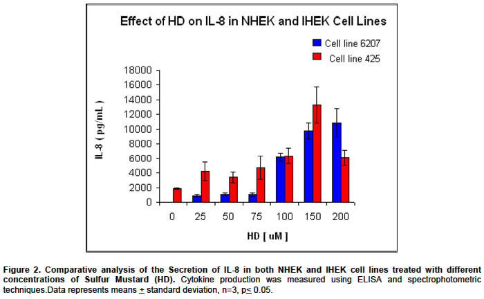
Code Number: ej04014 Financial support: Received October 7, 2003 / Code Number: ej04014 ABSTRACT Cytokines have been established as biomarkers to detect exposure of cells to chemical warfare agents such as sulfur mustard (2,2'-dichlorodiethyl sulfide, HD). In this study cultured normal and SV40 immortalized human epidermal keratinocyte (NHEK/IHEK) cells were compared as potential model systems to measure the efficacy of therapeutic drugs against HD. Immortalized human epidermal keratinocytes resemble their primary cell counterparts but have the advantage of being carried through long-term culture. Immortalized cells also provide consistency and durability and are less costly than primary keratinocytes. Immunoassay studies were performed to examine the response of these two cell lines to HD. We found that both normal and immortalized NHEKs secreted the pro-inflammatory mediator interleukin-8 (IL-8) when exposed to HD. However, a major difference was observed between the NHEK cell line 6207 and IHEK cell line 425. IHEK cell line 425 produced higher levels of Interleuken-8 then those of its normal counterpart cell line 6207. This observation is significant since therapeutic drugs such as ibuprofen, which depress cytokine production, may not allow these biomarkers to be detected efficiently in experimental analysis of certain NHEK cell lines. The fact that Il-8 production higher in cell line 425 cell makes this in vitro model a potential screening tool to study the efficacy of drugs that suppress production of cytokine markers. Keywords: chemical agent, human keratinocytes,interlukin-8, simian virus 40, sulfur mustard. Abbreviations: ARTICLE Epithelial cells are the initial target of a variety of toxic
chemicals, including the chemical warfare agent Sulfur Mustard (2,2'-dichlorodiethyl
sulfide, HD). The first time that HD was used as a chemical warfare agent was
on the 12th of July 1917, the German Army fired artillery rounds
filled with mustard at British troops near Exposure to sulfur mustard may be classified as high or low dose exposure. High dose exposure to HD leads to vesication in vivo and can be defined as exposure to a concentration above 50 µM in vitro.Concentrations between 50 µM and 100 µM lead to the disruption of cellular metabolic processes and the rapid onslaught of cell death occurs immediately above the 100 µM range. Since HD is a potent alkylating agent low dose exposure, which targets the DNA of the cell, causes long-term damage such as cutaneous carcinomas (Hurst and Smith, 2001). HD has been studied by using several different in vitro systems including; cultivated human fibroblast, cells derived from tumours and normal epithelial human keratinocytes (NHEK). These systems have provided insight into the metabolic and cellular reactions of chemical toxins, but are not without experimental challenges. Fibroblast and carcinogenic tissue may differ from their normal epithelial counterparts when exposed to chemical toxins. NHEK cells rapidly undergo senescence are expensive and come from a variety of different donors. Experiments which seek to study long-term/low-dose effects of toxic agents while using the NHEK in vitro system have encountered difficulties due to the cells limited growth potential. Cells transformed with Simian Virus 40 (SV40) have been established as immortalized cell cultures, that exhibit an indefinite growth potential (Steinberg and Defendi, 1979; Defendi et al. 1982; Steinberg and Defendi, 1983; Morris et al. 1985). These Immortalized human epithelial keratinocytes (IHEK) can be carried through long term culture, are cost effective and come from the same donor. SV40 transformed cell lines have been used as a tool for studying the effects of both mutagenic and nongenotoxic chemicals and therefore are an established model, which may be an ideal model for long-term/low-dose studies (Steinberg et al. 1999). Cytokines have been used as biomarkers to detect exposure of cells to chemical warfare agents such as HD (Arroyo et al. 1999). Therapeutic non-steroidal anti-inflammatory drugs (NASID) such as ibuprofen (IB), have been shownto inhibit the normal expression ofinflammatory cytokines. If cytokine are not detected efficiently in normal human keratinocyte cell lines then the use of an in vitro model where these cellular markers can be measured at toxicologically important concentrations must be established (Konstan et al. 1995; Stuhlmeier et al. 1999; Scheuren et al. 1998). SV40 immortalized human epidermal cells that produce higher concentrations of cytokines have been used as model cell lines to study the expression of cytokines such as IL-8 (Gerritsma et al. 1998; Chodosh et al. 2000; Petit-Frère et al. 2000; Walsh et al. 2001). We compared a normal cell line to a SV40 immortalized human epidermal keratinocyte cell line that secreted higher levels of the IL-8, so as to measure the efficacy of possible therapeutic intervention against the effects of low level exposure to HD. Experiments were conducted at physiologically and toxicologically significant concentrations of HD, which have been established as a HD concentration between the ranges of 25-100 µM (Hurst, and Smith, 2001). MATERIALS AND METHODS NHEK 6207 cell line which, in the 3rd serial passage, were obtained from Clonetics® and grown in Keratinocyte Growth Media (KGMTM). These cells are frozen in a cryoprotectant cocktail (growth medium, 10% v/v fetal bovine serum, and 10% v/v dimethylsulfoxide). The derivation, maintenance and growth properties of the SV40 immortalized human keratinocytes (IHEK) have been described previously (Steinberg and Defendi, 1979; Defendi et al. 1982; Steinberg and Defendi, 1983; Morris et al. 1985). Line 425 IHEKs at the 72nd and 80th serial passage were used in the experiments described here. Immortalized cells were grown in Dulbecco's Modified minimal essential medium (DMEM) supplemented with 10% fetal calf serum and 0.4 µg/ml hydrocortisone. Sulfur mustard (2,2'-dichlorodiethyl sulfide; HD) was acquired
from the U.S. Army Soldier and Biological Chemical Command ( Percent cell viability was determined using Propidium Iodide
(PI) and analyzed on a flow cytometer (FACSort, Becton Dickinson Immunocytometry
Systems, San Jose, CA, USA) using an argon laser at 488 nm. Mean percent viability
values and standard deviations were determined from three exposures per experimental
run as previously described (Clayson et al. 1993). Experiments
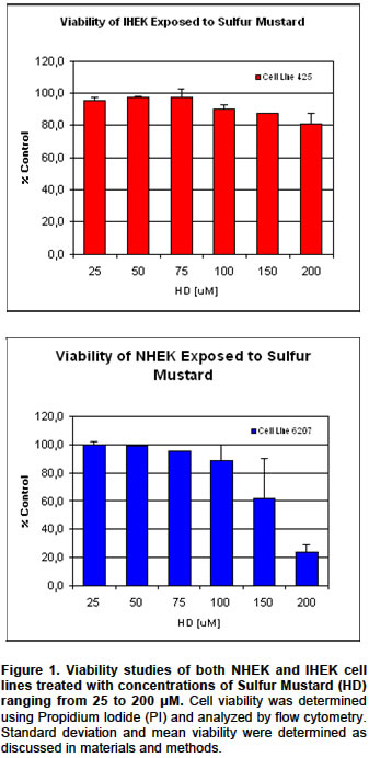
were run twice and data presented in (Figure 1). Viable
cells numbers was counted using a coulter counter (Coulter® Z1 Particle Counter, Enzyme-Linked Immunosorbent Assay (ELISA) Cytokine levels were measured using ELISA. Human IL-8 immunoassays
produced by Quantikineä (R&D
Systems, Inc., RESULTS Analysis of NHEK/IHEK cells exposed to different concentrations of HD revealed similar viability profiles at concentrations between 25 to 100µM, but differed at higher concentrations of sulfur mustard, above 100 µM (Figure 1). These observations are consistent with the fact that exposure of human epithelial cell lines exposed to a concentration of HD, that is between 1-100 µM is considered physiologically significant. At concentrations below 100 µM HD, when the secretion of IL- When normal cells were treated with ibuprofen there were no
detectible amounts of IL-8 either in the absence or presence of HD. But when
SV40 transformed cell line 425 was treated with the anti-inflammatory agent
ibuprofen the concentration levels of IL-8 went from 1.87 µg/mL to 0.319 µg/mL,
this is a 6-fold decrease in the production of the cytokine. We also observed
a decrease in the secretion IL- DISCUSSION The experiments shown here were undertaken to establish an in vitro human epithelial model to test the efficacy of drugs with potential to be used as countermeasures following exposure to vesicating agents, such as sulfur mustard. We compared two cellar models. The first was a commercially obtained normal human epithelial cell line (cell line 6207). The second was a SV40 transformed human epithelial cell line (cell line 425). Viability studies, using propidium iodide (PI) and flow cytometric analyses of cell integrity showed that at concentrations of sulfur mustard between 25 to 100 µM, the viability profiles of the normal and immortalized cells do not differ significantly. Since the viability of these cells are similar at concentrations of HD between 5 µM and 100 µM this may indicate that at both low dose and high dose exposures these cell lines physiologically react in a similarly fashion. However, at concentrations between 150 µM and 200 µM the mortality rate of NEK cells was higher than that of the IHEK cells. At concentrations of 150 µM to 200 µM of sulfur mustard cells are extensively damaged and start to die. This finding may be important in that experiments conducted at sulfur mustard concentrations of above 150 µM may use the immortalized cell lines as a model. Our results also showed that IHEKs behaved in the same manner
as NHEKs with regard to induction of IL-8 secretion in response to HD exposure.
Expression of interleukin- Results obtained in experiments using the non-steroidal anti-inflammatory
drug, ibuprofen, appeared to indicate that the amount of IL-8 secretion might
be an important factor in evaluating the efficacy of this drug in reducing
the inflammatory response. Studies conducted with NHEK in the presence of Ibuprofen
did not yield any detectible concentrations of IL-8 both in the cells exposed
and not exposed to sulfur mustard. In contrast, IL-8 levels in HD-treated IHEK
cells in replicate experiments were dramatically suppressed by IB treatment.
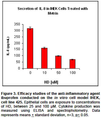
When SV40 transformed cell line 425 was treated with the anti-inflammatory
agent ibuprofen the concentration levels of IL-8 went from 1.87 µg/mL to 0.319 µg/mL,
this is a 6-fold decrease in the production of the cytokine. But it is important
to note that IL-8 concentrations were still detectable in the 425 cell line
but not in the normal cell line while using the same concentration of the prophylaxis
ibuprofen. We also observed a decrease in the secretion IL- These preliminary results support the idea that the SV40 transformed immortalized epidermal cells can be used as an useful, and inexpensive, alternative in vitro model system to test inflammatory processes stemming from cutaneous vesicant injury. REFERENCES
Note: Electronic Journal of Biotechnology is not responsible if on-line references cited on manuscripts are not available any more after the date of publication. Supported by UNESCO / MIRCEN network Copyright 2004 by Universidad Católica de Valparaíso -- Chile The following images related to this document are available:Photo images[ej04014f3.jpg] [ej04014f2.jpg] [ej04014f1.jpg] |
| |||||||||
