 |
| About Bioline | All Journals | Testimonials | Membership | News |
|
||||||
|
||||||
Electronic Journal of Biotechnology, Vol. 7, No.3, December, 2004, pp. 282-286 RESEARCH ARTICLE Plant regeneration of Anthurium andreanum cv Rubrun Teresa E. Vargas1, Alexander
Mejías2, Maira
Oropeza*3, Eva
de García4 Financial support: This work was supported by the Council for Scientific and Humanistic Development (CDCH), Central University of Venezuela grant PI 0333-4393-2002 to Maira Oropeza. Code Number: ej04032 To establish an efficient regeneration system for Anthurium andreanum cv Rubrun, seeds from plant spadixes were germinated on a medium supplemented with 2.2 mM BA. After 2 weeks, 74% of the seeds germinated and four weeks later, micro-cuttings from these plantlets were subcultured on a medium containing 4.4 mM BA and 0.05 mM NAA. On average, 3.6 shoots per explant were obtained. Four weeks old in vitro plants from germinated seeds and the plantlets obtained from micro-cuttings, showed callus proliferation at the stem base. These tissues were subcultured on a medium supplemented with 8.9 mM BA and 2.7 mM NAA. After 6 weeks of culture, about 43.8 plantlets per square cm of callus were obtained. Anatomical studies showed the organogenic nature of these calli. Anthurium andreanum plants regenerated by organogenesis were transferred to pots and a rate of 80% of plant acclimatization was obtained. Keywords: Callus, micropropagation, organogenesis. Abbreviations: ARTICLE The Anthurium genus comprises about 1500 tropical species which are important ornamental plants and are normally propagated by seed (Dufour and Guerin, 2003). Vegetative propagation methods applied to these plants have not shown good results and tissue culture techniques appear as an alternative to increase the production (Pierik et al. 1974, Chen et al. 1997). Plant regeneration of Anthurium andreanum has been achieved through adventitious shoots formation from callus (Pierik et al. 1974; Pierik and Steegmans, 1976) and direct shoot regeneration from lamina explants (Martin et al. 2003). Teng (1997) established the fact that in liquid or raft cultures most adventitious Anthurium shoots regenerated singly or in loose aggregates, which is an advantage over solid cultures. Geier (1986) concluded that plant age and plant genotype influence plant regeneration of Anthurium andreanum, and analyzed the influence of NH4NO3 on callus and shoots formation from young leaf tissues. Kuehnle and Sugii (1991) established a regeneration system from leaves and petioles of Anthurium Hawaiian cultivars through callus tissue cultures, and Kunisaki (1980) established the micropropagation of Anthurium from axillary buds. Chen et al. (1997) regenerated Anthurium andreanum plants from roots explants. Reliable proliferation of callus and subsequent plant regeneration are important for massive plant propagation, studies on genetic transformation and development of transgenic plants with new trails. The improvement of the micropropagation system for this species could be important for its commercial applicability. This paper describes the preliminary results for the establishment of an alternative method for regeneration of Anthurium andreanum cv. Rubrun plants from callus tissue through organogenesis. Micropropagation of this plant species after seed germination is also described.
MATERIALS AND METHODS
Explants
were obtained from plants of A. andreanum cv Rubrun germinated from
seeds. The fruits were separated from spadixes and sterilized for 15 min
in 3% NaOCl and then rinsed three times with sterile water for 30 min, following
the protocol established by Pierik et al. (1974), modified
as follows: one hundred seeds were isolated and sterilized for 20 min in
1% NaOCl, and afterwards they were washed two times with sterile water for
30 min. Two media were tested for seed germination. They shared a basal composition
of Murashige and Skoog, (1962), supplemented with 1.2 mM
thiamin, Micropropagation of Anthurium andreanum After
4 weeks, we used one of the plants germinated from seeds as source of explants.
Four micro-cuttings were inoculated on Murashige and Skoog,
(1962) medium supplemented with 4.4 mM BA and 0.05 mM NAA and incubated
under continuous fluorescent light (50 mE. m-2.s-1)
at At the
stem base of the 8 weeks-old plants originated from micro-cuttings, a proliferation
of callus tissue was observed. Twenty segments of approximately 1 x Plantlets originated from callus tissue were maintained on the same medium during 4 months and then were transferred to Murashige and Skoog, (1962) medium without hormones for three months. Later, these 7 months old plants were removed from the culture tubes. The roots were washed in tap water and the plants were transferred to pots containing a mixture of soil and organic humus (1:1). The plants were kept on chambers with high relative humidity and low light intensity. One month later, when the plants shown growth, they were transferred to the greenhouse. For the histological observations the tissues were fixed in FAA (40% formaldehyde, 10% glacial acetic acid, 50% ethanol) for at least two days, followed by dehydration in series of tert-butyl-ethanol and ethanol (De García and Martínez, 1995). Then they were embedded in paraplast for sectioning in a rotatory microtome (10 - 12 mm) followed by staining with safranine and fast green. The observations and photomicrograph were done with a Wild Heerbrugg optical microscope model M20EB, equipped with a camera.
RESULTS
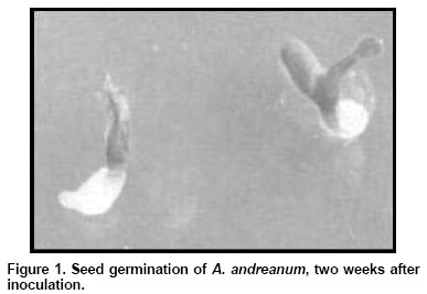
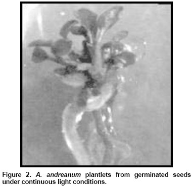
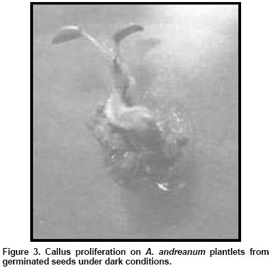
In vitro seed germination of Anthurium andreanum After
one week, 74% of the seeds cultivated under continuous light conditions were
germinated in comparison with 30% of germination showed by seeds cultivated
on Murashige and Skoog, (1962) medium with BA incubated
in darkness. One week later the radicle emerged and shoot developed under
continuous light (Figure 1). On the contrary, germinating
seeds cultivated on darkness did not show development of shoots and radicle
at this time. The germinated seeds were all transferred to a medium without
hormones under light conditions. Two weeks later, the plantlets previously
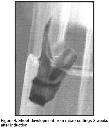
developed under light conditions were Micropropagation of Anthurium andreanum plants from micro - cuttings After
two weeks, buds from micro-cuttings cultivated on Murashige
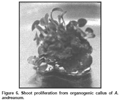
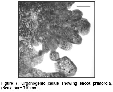
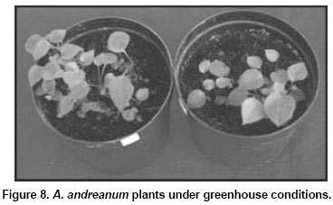
and Skoog, (1962) medium enlarged Plant regeneration through callus culture Callus tissues originated at the stem base of A. andreanum plantlets obtained from micro - cuttings culture, were sub-cultured and 6 weeks later these green and compact calli had increased their size twice. We obtained an average of 43.8 plantlets per callus fragment (Figure 6). Anatomical studies showed that these green calli had organogenic potential (Figure 7) showing numerous shoot primordia. Under the acclimatization conditions used in this work, the success rate was 80%. The acclimatized plants showed the same morphology than the Anthurium andreanum plants used as seeds source (Figure 8).
DISCUSSION
An efficient
regeneration system for A. andreanum from micro - cuttings and from
callus tissue was achieved in this research. Previous reports have shown
that the vegetative propagation of several Anthurium species is a
very difficult task (Pierik et al. 1974; Pierik
and Steegmans, 1976; Hamidah et al. 1997), moreover,
the application of culture techniques, have been also difficult to establish
because of the high contamination indexes observed in these culture (Brunner
et al. 1995). We showed here that using plants from germinated seeds
permitted to eliminate the contamination problems we previously encountered
using explants from plants grown in the greenhouse. Our results show that
there are high germination percentages of A. andreanum seeds, under
continuous light conditions. Plantlets issued from seeds and callus formed
at the basal zones of the micropropagated plants, allowed to set up alternative
regeneration systems for A. andreanum. Kuehnle and Sugii
(1991) obtained embryogenic callus from plant spadixes and organogenic
callus from leaf explants of several Anthurium hybrids. They maintained
long-term cultures of callus from Note: Electronic Journal of Biotechnology is not responsible if on-line references cited on manuscripts are not available any more after the date of publication. Copyright 2004 by Universidad Católica de Valparaíso -- Chile The following images related to this document are available:Photo images[ej04032f2.jpg] [ej04032f4.jpg] [ej04032f8.jpg] [ej04032f5.jpg] [ej04032f7.jpg] [ej04032f3.jpg] [ej04032f6.jpg] [ej04032f1.jpg] |
| |||||||||
