 |
| About Bioline | All Journals | Testimonials | Membership | News |
|
||||||
|
||||||
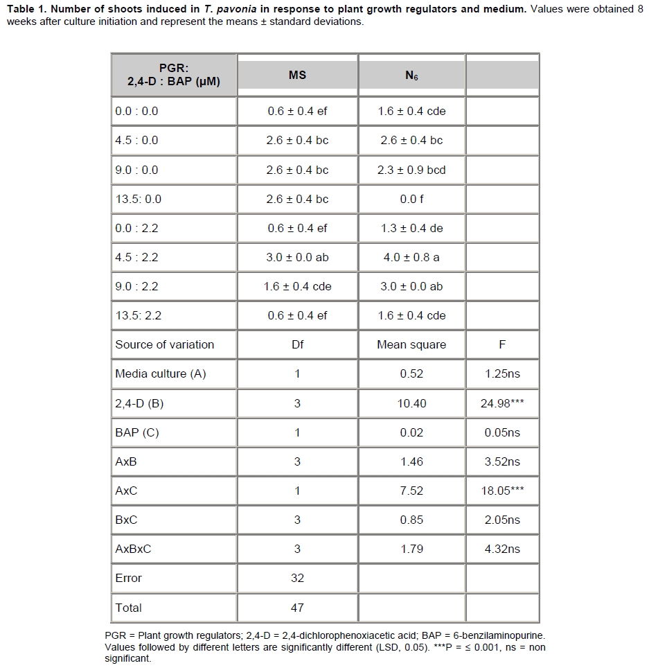
Electronic Journal of Biotechnology, Vol. 13, No. 1, Jan, 2010 Research Article In vitro regeneration and genetic fidelity of Tigridia pavonia (L.f.) DC. José Luis Piña-Escutia1 , Luis Miguel Vázquez-García2, Amaury Martín Arzate-Fernández*3 1Centro de Investigación y Estudios Avanzados en
Fitomejoramiento,
Facultad de Ciencias Agrícolas,
Universidad Autónoma del Estado de México,
Toluca, Estado de México, México Financial support: We are grateful to Consejo Nacional de Ciencia y Tecnología (CONACYT) from México for the scholarship give to JLPE (2005-183935), and to the RED TIGRIDIA (SINAREFI) for covering the publishing payment. Received January 12, 2009 / Accepted September 8, 2009 Code Number: ej10007 Abstract Plants of Tigridia pavonia (L.f.) DC were regenerated from twin-scaling explants cultured on Murashige and Skoog and N6 basal medium. The highest formation of shoots per responding explant was obtained on N6 medium supplemented with 4.5 µM 2,4-dichlorophenoxy acetic acid in combination with 2.2 µM benzylaminopurine. Shoots rooted readily on N6 basal medium supplemented with 1 g l-1 activated charcoal and 2.6 µM naphtalenacetic acid. The rooted shoots achieved 100% survival. Inter Simple Sequence Repeat analysis was carried out to check for possible genetic alterations in plants obtained after two consecutive subcultures. The results revealed that the recovered plants did not exhibit any type of polymorphism. Keywords: genetic integrity, micropropagation, oceloxóchitl.
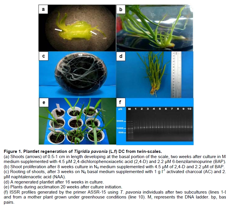
Tigridia pavonia (L.f.) DC., also known as tiger flower or oceloxóchitl, is a native bulbous plant of Mexico and it was used by the Aztecs as an ornamental, and for food and medicinal purposes. Although in Mexico there is no record of its commercial production, it is commercialized as garden plant in Europe, Asia and Australia where is known for its flowering longevity of only 10-14 hrs. This species have been strongly extracted, and its phytogenetic germplasm could be lost (Vázquez-García et al. 2001). T. pavonia is usually propagated in one of two ways: by seed or by bulb. The use of seeds is not a practical propagation method because of the semi-compatibility and low germination rate. Moreover, to obtain stock bulbs, from two to three years is required because of the slow growth of the bulbs until flowering (Vázquez-García et al. 2001). Tissue culture can be employed as an alternative to conventional methods of vegetative propagation with the goal of enhancing the rate of multiplication of desired genotypes (Paek and Murthy, 2002). The technique of twin-scaling has been effective for tissue culture propagation of Iris hollandica (Fidalgo et al. 2005) and other bulbous species, including Eucrosia stricklandii (Colque et al. 2002), Narcissus (Santos et al. 2002) and Sternbergia clusiana (Sawsan and Fattash, 2005). Despite the advantages of in vitro propagation, factors such as growth regulators, time of culture and media composition appear to be capable of inducing in vitro genetic variability in the regenerated plantlets (Silvarolla, 1992). Several approaches have been applied for identification of variants among micropropagated plants including: phenotypic variation (Kancherla and Bhalla, 2003), karyotypic analysis of chromosomes (Bennici et al. 2004), and biochemical analysis (Punja et al. 2004). A major disadvantage of these techniques is the availability of only limited numbers of informative markers since the number of assays to detect them is relatively low. Therefore, the percentage coverage of the genome is inadequate to make a comprehensive study of genetic diversity. Furthermore, the markers can be influenced by environmental factors, and the expression differences can cause confusion in the interpretation of the results (Vicente and Fulton, 2003). Inter Simple Sequence Repeat (ISSR) markers, which use anchored primers to amplify simple sequence repeats without the requirement for prior sequence information, is a very simple, fast, cost-effective, highly discriminative and reliable technique (Reedy et al. 2002), and they have been successfully applied to detect the genetic similarities or dissimilarities in micropropagated material in Prunus dulcis (Martins et al. 2004), Tulipa spp. (Podwyszynska et al. 2006), Musa spp. (Lakshmanan et al. 2007), and Vitex negundo (Ahmad et al. 2008). To date, there have been no reports on in vitro propagation of T. pavonia. Here, we describe an efficient protocol for direct plant regeneration from twin-scale explants of T. pavonia along with the ISSR analysis to determine the genetic fidelity of the regenerated plants. Materials and Methods Plant material and disinfection T. pavonia (L.f.) DC bulbs were obtained from plants grown in a greenhouse, and were used as source material to initiate in vitro cultures. The outer scales, one-third of the upper part and the dry remains of the previous root disks of the bulbs were removed, and the bulbs were placed in a flask with 10 ml of antibacterial soap (Reckitt Benckiser Inc. USA) and rinsed in running tap water for 30 min. Bulbs were then soaked for 1 hr in a solution containing 1% (w/v) Benomyl (metil-1-butilcarbomoil-2-bencinidazol-carbamate a.i. 50% w/w) and 1% (w/v) Agry-Gent (gentamycin sulphate a.i. 2% w/w and Oxytetracyclin chloride a.i. 6% w/w). Afterwards, in a laminar flow cabinet, bulbs were surface disinfected with 70% ethanol for 2 min, then immersed in 5% sodium hypochlorite for 10 min followed by 5% calcium hypochlorite for 15 min; finally, they were washed three times with distilled sterilized water. Each disinfected bulb was longitudinally sectioned to obtain explants formed by segments of twin-scales, 2-3 mm wide and 6-8 mm length, joined by a thick segment (2-3 mm) of basal tissue. The explants were cultured upright with the basal plate tissue inserted into the culture medium. Medium culture and shoot induction For shoot induction, two basal media: MS (Murashige and Skoog, 1962) and N6 (Chu et al. 1975) and 0 and 2.2 µM concentrations of 6-benzylaminopurine (BAP) and 0, 4.5, 9 and 13.5 µM 2,4-dichlorophenoxiacetic acid (2,4-D) in all possible combinations, were tested. The basal media were supplemented with 30 g l-1 sucrose and 1 g l-1 activated charcoal. All media were adjusted to a pH of 5.7 ± 0.1 and then 2.5 g l-1 gelrite was added before autoclaving at 121ºC and 1.1 kg/cm2 for 20 min. Eight explants were placed on each sterile Petri dish (90 mm diameter) containing 20 ml of medium, then sealed with ParafilmTM. Cultures were incubated at 25 ± 1ºC under cool-white fluorescent tubes with a light intensity of 34 ± 5 µmol m-2 s-1 and 16 hrs photoperiod. Explants were subcultured every 30 days. Shoots of 4-6 cm in length were separated from the original explant, and subcultured in glass flasks (500 ml, covered with aluminum plugs) on N6 basal medium supplemented with 1 g l-1 activated charcoal and 2.6 µM naphtalenacetic acid (NAA) to induce rooting. The incubation conditions and light intensity were the same as stated above. Plants with a mean height of 15 cm and a well-developed root system were removed from glass flasks and transferred to ex vitro conditions. For this purpose, they were rinsed free of tissue medium culture with distilled water, and potted in plastic pots (25 cm3) containing a sterilized mixture (1:1) of compost and agrolita (DICALITE®). Clear polyethylene bags (300 cm2) were inverted over the pots and secured with rubber bands. The bags were ventilated by removing the rubber bands after 2 weeks; bags were removed 1 week later and the plants were then transferred to a greenhouse. To evaluate the effect of each treatment on shoot induction, a factorial analysis and LSD test were performed using the data of number of shoots per explant formed 8 weeks after culture initiation. The treatments were arranged in a completely randomized design. Data were analyzed with Statistical Analysis System software version 8.0 (SAS Institute, 1999). DNA extraction and ISSR analysis DNA was extracted from 100 mg of fresh leaves derived from one to four shoots from each treatment after two subcultures (30 d each) and from a mother plant grown under greenhouse conditions. DNA extraction was carried out according to Arzate-Fernández et al. (2005). The DNA samples were stored at -20ºC until be used. Five anchored simple sequence repeat primers (3'-ASSR02, 3'-ASSR15, 3'-ASSR20, 3'-ASSR29 and 3'-ASSR35) (Yamagishi et al. 2002) were used for the Polymerase Chain Reaction (PCR). Amplification reactions were performed in 10 µl containing 1 µl of 10X PCR ammonium buffer (1.5 mM MgCl2), 0.5 µl of MgCl2 (25 mM), 1 µl of dNTPs (10 mM) (APPLIED BIOSYSTEMS®), 1 µl of the primer (20 µM) (INVITROGENTM), 0.1 U of Taq DNA polimerase (MERCURY REAGENTSTM) and 1 µl of DNA (10 ng). DNA amplification was performed in a thermal Mastercycler gradient (EPPENDORF®). The cycles for the primers 3'-ASSR02 and 3'-ASSR15 were according to Yamagishi et al. (2002) and Arzate-Fernández et al. (2005) with modifications comprising the following: an initial cycle of 9 min at 94ºC, 1 min at 46ºC and 1 min at 72ºC; then 45 cycles of 1 min at 94ºC, 1 min at 46ºC and 1 min at 72ºC followed by one final extension cycle of 9 min at 94ºC, 1 min at 46ºC and 10 min at 72ºC. The cycles for the primers 3'-ASSR20, 3'-ASSR29 and 3'-ASSR35 were according to Yamagishi et al. (2002). Amplified products were separated by 1% agarose gel (w/v) (SIGMA®) electrophoresis at 100 V. The gel was stained with 3 µl of ethidium bromide and the DNA bands were photographed under ultraviolet light using a UV Transilluminator BioDoc-It Imaging System (UVPTM). The sizes of the amplification products were estimated with a DNA ladder (100 bp to 3.0 kb) (FERMENTASTM). The ISSR profiles were analyzed on the basis of the presence or absence of individual ISSR bands. Results and Discussion The response of explants cultured on MS and N6 medium culture with 2,4-D and BAP on shoot induction of T. pavonia is shown in Table 1. After two weeks of culture, shoots arose from the basal portion of the bulb scales (Figure 1a) in some treatments. In those responding treatments, the original explant maintained its organogenic capacity for more than 12 weeks; during which adventitious shoot proliferation was observed. The period for shoot induction reported here, is faster in comparison with other bulbous species such as Eucrosia stricklandii, where a response was obtained in four weeks (Colque et al. 2002), and Narcissus asturiensis, where it occurred at eight weeks (Santos et al. 2002). In this study, up to 90% of the explants developed shoots and the number of shoots per explant varied between 0 and 4 (Table 1). Although the number of shoots per explants was low, it was similar to shoot numbers reported in other bulbous plants; for example, in Eucumis autumnalis 1.0 to 3.7 shoots per explant were obtained (Ault, 1995), and in Eucrosia stricklandii 0.7 to 2.2 shoots per explant was observed (Colque et al. 2002). The effect of 2,4-D and of the interaction of medium and BAP on shoot induction, after eight weeks, was highly significant (Table 1). It has been reported that auxin is an effective PGR for shoot induction and in combination with a cytokinin, is essential for shoot induction (Paek and Murthy, 2002; Santos et al. 2002). In this study, the combination of 2,4-D with BAP stimulated shoot induction (Figure 1b). Our results agree with those reported by Boltenkov and Zarembo (2005) in Iris ensata, and Anbari et al. (2007) in Narcissus papyraceus, who reported that combinations of 2,4-D and BAP were necessary for shoot induction. The combination of low levels of 2,4-D (4.5 µM) with BAP (2.2 µM), produced three and four shoots per explant in MS and N6 media respectively (Table 1): but in high levels of 2,4-D (13.5 µM) with BAP (2.2 µM), the response was minor (0.6 and 1.6 shoots, respectively) and the shoots formed in these levels were smaller to those obtained in low levels. Similar results were observed by Aftab et al. (2008) who reported sporadic single shoot regeneration in Gladiolus hybridus using high levels of 2,4-D (9 µM). In contrast, Mata-Rosas et al. (2006) obtained the best shoot formation in Magnolia dealbata using low concentrations of 2,4-D (0.5 µM). The results of this study, and the above mentioned work, suggest that low levels of 2,4-D in combination with BA, are enough for in vitro shoot induction, but the use of 2,4-D at high levels affects the normal development of plant tissues, which was also observed by Filippi et al. (2001) and Steinitz et al. (2003) in Musa spp and Capsicum annum, respectively. Although the effect of MS and N6 on shoot induction was not significant, the rate of shoot induction was slightly higher in N6 medium in comparison with the MS medium (Table 1). MS medium has been reported as one of the best medium for shoot induction (Colque et al. 2002; Santos et al. 2002; Sawsan and Fattash, 2005) while N6 medium has been used in few species, for the same purpose. MS medium differs significantly from N6 medium with respect to the content of ammonium and nitrate ions, and also to a lesser degree with the content of phosphate ions. Given that both elements (nitrogen and phosphorus) are consumed quickly by the explants cultivated in vitro, they can be limiting factors in tissue culture growth (Ramage and Williams, 2002). Also, it has been observed that high ammonium levels have a negative effect on growth and morphogenesis. Considering this, it is possible that the highest shoot formation response observed in the N6 medium in this study, may have been the result of the higher quantity of phosphorous and the smaller quantity of ammonium present in the N6 medium in comparison with MS medium. It has been reported that germination of T. pavonia seeds is very low, while its propagation by bulb can produce four bulbs with same number of plants in a two to three year period (Vázquez-García et al. 2001). In contrast, the in vitro regeneration protocol described in this study may produce 48 plants from one bulb in a period of five months (an average of 12 scales per bulb and four plantlets per scale). This is the first report for in vitro culture of T. pavonia and the procedure described is an effective regeneration protocol, although further experiments on the explant size will be required to improve shoot induction. It has been reported that the AC improved shoot formation, plant recovery and rooting (Arzate-Fernández et al. 2007), and that low NAA concentrations (0.5-2.5 µM) are optimal for the root induction (Colque et al. 2002; Aftab et al. 2008; Torabi-Giglou and Hajieghrari, 2008). In this study, the addition of AC and NAA showed a positive effect because all adventitious plantlets (4-6 cm in length) continued developing on N6 basal medium supplemented with 1 g l-1 AC and 2.6 µM NAA, and after 3 weeks culture, roots formed (Figure 1c). Our results are in agreement with Ault (1995), and Mata-Rosas et al. (2006) who also reported beneficial effects of AC and NAA on in vitro root stimulation in Eucrosia autumnalis and Magnolia dealbata respectively. Pospíšilová et al. (1999) mentioned that, through in vitro culture, the plantlets can develop morphological, anatomical and physiological abnormalities. Also, regenerated plantlets often die during transfer from in vitro to the ex vitro conditions. In contrast, in this study, sixteen weeks after culture initiation, more than 100 regenerated plants (10-15 cm in length) with roots (4-6 cm) and 2-4 green leaves were obtained (Figure 1d) and these were established with a mixture of compost and agrolita and transferred to a greenhouse with a 100% success rate (Figure 1e). It is possible that the development of complete plants and the ex vitro handling protocol may have contributed to the adaptation success. Genetic uniformity is one of the most important prerequisites for the successful micropropagation of any crop species. Nevertheless, a major problem encountered in cells grown in vitro is the occurance of genetic variation. Although the origin of this variation is unclear, it is considered that there are two main factors: an intrinsic factor, which largely depends on the genetic stability of the explants, and an extrinsic factor, depending on culture media and particularly growth regulators (in particular 2,4-D) (Smith, 1998). The ISSR technique has been used successfully for analyzing the genetic stability of species propagated through in vitro shoot formation (Martins et al. 2004; Podwyszynska et al. 2006; Lakshmanan et al. 2007; Ahmad et al. 2008). In this study, ISSR analysis did not reveal any type of polymorphism between randomly selected in vitro plants of T. pavonia and the mother plant, indicating the clonal nature of progeny (Figure 1f). This observation constitutes an advantage for the in vitro regeneration procedure because it minimizes the risk of genetic variation. The five ASSR primers tested in this study generated 21 scorable band classes (all monomorphic) ranging in size from 500 bp (ASSR-20) to 1700 bp (ASSR-35). The number of bands for each primer was from 3 (ASSR-02 and ASSR-15), 4 (ASSR-29), 5 (ASSR-35) to 6 (ASSR-20) with an average of 4.2 bands per ASSR primer. An example of PCR amplification products obtained with the primer ASSR-15 is given in Figure 1f. This findings agree with those reported by others (Lakshmanan et al. 2007; Ahmad et al. 2008; Bhatia et al. 2009) who have pointed out the efficacy of 3'-anchored primers for giving a higher number of clearly definable bands and being better than other markers such as RAPDs in the assessment of clonal fidelity of micropropagated plants (Martins et al. 2004). Concluding Remarks The best conditions for in vitro shoot induction using twin-scale explants of T. pavonia was the treatment with 4.5 µM 2,4-D and 2.2 µM BAP in N6 medium supplemented with 1 g l-1 of activated charcoal, and resulted in four shoots per explant. Thus, 48 plants can be produced from one bulb within five months of culture initiation. N6 medium supplemented with 1 g l-1 activated charcoal and 2.6 µM NAA can be used to induce rooting. The protocol described is an effective regeneration procedure for T. pavonia because no somaclonal variation was observed among the regenerated plants based on ISSR analysis. References
Note: Electronic Journal of Biotechnology is not responsible if on-line references cited on manuscripts are not available any more after the date of publication. Supported by UNESCO / MIRCEN network Copyright © 2010 by Pontificia Universidad Católica de Valparaíso -- Chile The following images related to this document are available:Photo images[ej10007t1.jpg] [ej10007f1.jpg] |
| |||||||||
