 |
| About Bioline | All Journals | Testimonials | Membership | News |
|
||||||
|
||||||
Electronic Journal of Biotechnology, Vol. 13, No. 4, July 15, 2010 Rapid multiplication and in vitro production of leaf biomass in Kaempferia galanga through tissue culture Reena Parida1, Sujata Mohanty2, Ananya Kuanar3, Sanghamitra Nayak*4 1, 2, 3Centre of
Biotechnology
Siksha ‘O’Anusandhan University
PO- Khandagiri, Bhubaneswar-751030
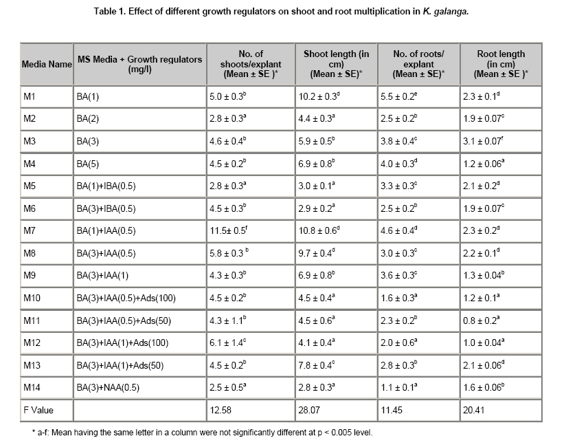
Orissa, India Code Number: ej10037 An efficient protocol has been established for rapid multiplication and in vitro production of leaf biomass in Kaempferia galanga L, a rare medicinal plant. Different plant growth regulators like Benzyladenine (BA), Indoleacetic acid (IAA), Indolebutyric acid (IBA), Napthaleneacetic acid (NAA) and adenine sulphates (Ads) have been tried for induction of multiple shoots using lateral bud of rhizome as explants. The highest rate of shoot multiplication (11.5 ± 0.6) shoot/explant as well as leaf biomass production (7.4 ± 0.3) gram/explant was observed on Murashige and Skoog medium supplemented with Benzyladenine (1 mg/l) and Indoleacetic acid (0.5 mg/l). Data of shoot multiplication and leaf biomass production were statistically analysed. Upon excission of leaves after 2 months of culture under sterile condition, the base of each plantlet was transferred to fresh media which could produce the same leaf biomass within another 2 months in a 50 ml culture tube containing 20 ml and 250 ml conical flasks containing 30 ml Murashige and Skoog medium. The rate of multiplication and leaf biomass production remained unchanged in subsequent subcultures. The regenerated plantlets were acclimatized in greenhouse and subsequently transferred to the field. Survival rate of the plantlets under ex vitro condition was 95 percent. Genetic fidelity of the regenerants was confirmed using random amplified polymorphic DNA (RAPD) marker. The protocol could be commercially utilized for large scale production of true-to-type plantlets and as an alternative method of leaf biomass production in Kaempferia galanga. Keywords: growth regulators, Kaempferia, in vitro propagation, leaf biomass.
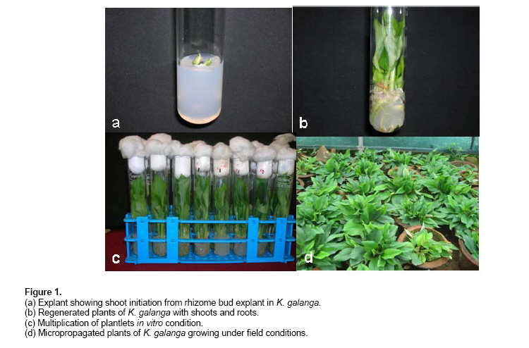
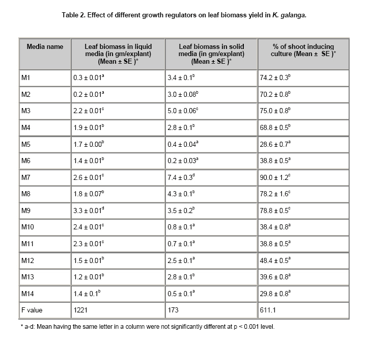
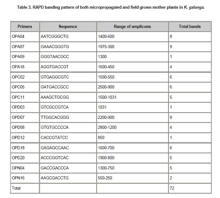
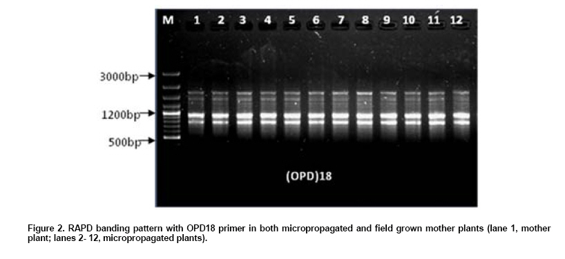
Kaempferia galanga L. (K. galanga) is a rare Indian medicinal herb of family Zingiberaceae. It is distributed in the tropics and subtropics of Asia and Africa. The plant is extensively used in preparation of ayurvedic drugs, in perfumery, cosmetics and as spice ingredients (Rahman et al. 2004). The plant is used for treatment of diarrhea, migraine and it increases energy to overcome exhaustion. Leaves of K. galanga are used in flavouring foodstuffs, hair tonics, mouth washes and also in cosmetic industries. The leaves possess antioxidant, antinociceptive and anti-inflammatory activities that help in treatment of mouth ulcers and headaches (Sulaiman et al. 2008; Chan et al. 2009). The poultices of leaves as well as the tea of leaves are used for curing sore throat, rheumatism, swellings and eye infections, whereas the leaf infussions of K.galanga can be used as a beneficial drink for pregnant women (Rahman et al. 2004; Sulaiman et al. 2008). The ashes of leaves are rubbed on swollen breasts after childbirth but fresh leaves are chewed for relieving coughs (Sulaiman et al. 2008). The plant is wild in nature and exhibits poor natural regeneration by rhizomes thus reaching the threshold of being recognized as an endangered species (Shirin et al. 2000). Large scale deforestation and over exploitation further reduces the plant population. K. galanga is rarely cultivated at some places for medicinal purposes. Conventional propagation of Kaempferia by the splitting of rhizome is not sufficiently rapid to meet the need of planting materials and leaves, and it takes years to build up for the commercial quantities. In the recent years micropropagation techniques are being profitably used to overcome the present demand of aromatic and medicinal plants (Rahman et al. 2004). Tissue culture techniques hold promise in this context for rapid multiplication and in vitro production of leaf biomass in K. galanga. In vitro production of leaf biomass has also been reported in few other species having commercial implications (Sharada et al. 2007; Kuanar et al. 2009). The genetic stability of tissue cultured plants is of utmost importance for commercial utilization of the technique for large scale production of true-to-type plants of desired genotype (Mohanty et al. 2008). Out of several molecular markers used for the assessment of the genetic stability, random amplified polymorphic DNA (RAPD) analysis is the simplest, cheapest and quickest method for determining the genetic fidelity of in vitro grown plants as reported in many species (Rout et al. 2002; Martins et al. 2004; Venkatachalam et al. 2007). The few reports so far available on tissue culture studies in K. galanga (Vincent et al. 1992; Shirin et al. 2000; Rahman et al. 2005) do not include any work on in vitro production of extractable leaf biomass for commercial purposes and assessment of genetic stability of micropropagated plants. The present paper deals with a suitable protocol for rapid propagation of genetically uniform plants and a novel method of in vitro production of leaf biomass in K. galanga for the first time. Materials and MethodsThe rhizomes of K. galanga were collected from the High Altitude Research Station, Pottangi of Orissa and were grown in the medicinal plant garden of Center Of Biotechnology, Siksha ‘O’Anusandhan University, Bhubaneswar. The rhizome sprouted during monsoon (June). The young sprouting buds from rhizome of 10 plants of K. galanga were used as explants for culture initiation. The explants were washed under running tap water followed by a neutral liquid detergent (Extran, Merck) for 3 to 5 min. The explants were then washed repeatedly with distilled water and finally surface sterilized with 0.1% mercuric chloride for 8 to 10 min in a laminar flow cabinet. Surface sterilized explants were washed 3 to 4 times with autoclaved water aseptically to remove any trace of mercuric chloride prior to cultivation. In vitro plant multiplication and cultural conditions Explants were inoculated to the basal medium of MS (Murashige and Skoog, 1962) with 1-5 mg/l of BA individually or with varying combination of 0.5 mg/l of IBA, 0.5-1.0 mg/l of IAA, 0.5 mg/l of NAA and 50-100 mg/l of Ads (Table 1). The sucrose amount in the media was 30 gm/l. There were ten replicas for each treatment and the experiment was repeated thrice. The medium pH was adjusted to 5.7 before adding agar and media were autoclaved at 121ºC and 105 kg/cm2 of pressure for 20 min. After sterilization of media the explants were transferred into the medium. Culture tubes (50 ml) and conical flasks (250 ml) containing explants were kept under white fluorescent light with 50 µmol/(m2s2) light intensity. Twenty and thirty ml media were dispensed respectively to culture tubes and conical flasks. Percentage of response of explants with different hormonal combinations was observed time to time and sub culturing was done at two month interval. Estimation of in vitro leaf biomass yield The in vitro grown plants were transferred to MS basal medium supplemented with varying combinations and concentrations of BA, IAA, NAA, Ads. After 8 weeks of growth, leaf biomass in solid and liquid culture, length of shoots and roots, number of shoots and roots were measured from 15 different culture tubes for each treatment. The leaf biomass yield in 250 ml flasks was also calculated after two months. After the first in vitro collection of leaves, shoot bases were separately transferred to culture tubes and flasks for production of another batch of leaf biomass. The process was repeated throughout the year. Establishment of plants in soil In vitro grown plants having well developed shoots and roots were washed gently under running tap water to remove agar from roots and planted in earthen pots containing soil, cowdung and sand mixture in 1:1:1 ratio. The plantlets were kept in greenhouse for acclimatization for 30 days and then transferred to normal field condition for growth until maturity. The percentage of survival was noted. The genomic DNA of K.galanga was isolated by following the protocol of Doyle and Doyle (1987) with some modification from both in vitro and ex vitro grown mother plants. The crude DNA was purified by adding RNAse A (60 µg for 1 ml of crude DNA solution) followed by washing with phenol:chloroform:isoamyl alcohol (24:24:1) and subsequently with chloroform:isoamyl alcohol (24:1). Then centrifugation was done and the upper aqueous layer was separated and mixed with 1/10th volume of 3M-sodium acetate. DNA was precipitated by adding 2.5 volume of chilled absolute ethanol; pellet was dried in vacuum and dissolved in Tris10EDTA1 buffer. Quantification of purified DNA was done by analyzing the purified DNA in 0.8% of agarose gel with a known uncut lambda DNA as standard. Purified DNA samples were then diluted in TE buffer to 25 ng/µl for RAPD-PCR amplification. For RAPD analysis a total of 18 random primers were used out of which 15 random decamer primers (Operon Tech, Almeda, USA) from A, C, D and N series (OPA 04, 07, 09, 18; OPC02, 05, 11; OPD03, 07, 08, 12, 18, 20 and OPN04, 16) were selected on the basis of clarity of banding patterns. The RAPD analysis was performed as per the method of Williams et al. (1990). PCR was performed in a volume of 25 µl containing 25 ng of genomic DNA as template, 2.5 ml of 10x assay buffer (100 mM Tris hydrogen chloride, pH 8.3, 500 mM potassium chloride and 0.1% gelatin), 1.5 mM magnesium chloride, 200 µM dNTPs (Bangalore Genei, India), 0.5 unit (U) of Taq polymerase (Bangalore Genei, India) and 15 ng of RAPD primer (Bangalore Genei, India). The amplification was carried out in a thermal cycler (Gene Amp PCR system 9700, Applied Biosystems, CA, USA). In PCR, the first step was performed at initial temperature of 94ºC for 5 min for complete denaturation of the template DNA. The second step consisted of 42 cycles having three ranges of temperature, i.e. at 92ºC for 1 min for denaturation of template DNA, at 37ºC for 1 min for primer annealing at 72ºC for 2 min for primer extension followed by running the samples at 72ºC for 7 min for complete polymerization. The PCR products obtained from RAPD were analyzed in 1.5% agarose gel stained with ethidium bromide (0.5 µg/µl) of gel solution in TAE buffer (40 mM Tris base, 20 mM sodium acetate, glacial acetic acid to pH 7.2) for 3 hrs at 60 volts. The size of the amplicons were estimated using 100 bp DNA ladder plus or DNA ladder mix (MBI Fermentas, Lithuania) as standard. DNA fingerprints were visualized under UV light and photographed using Gel Documenting system (Bio-Rad, USA). RAPD analysis using each primer was repeated at least twice to establish reproducibility of banding pattern of different DNA samples of K. galanga. Data were subjected to analysis of variance for a factorial experiment. Critical differences (CD) were calculated to determine the statistical significance of different treatment means. Results and DiscussionIn vitro shoot multiplication and leaf biomass yield The rhizome buds of conventionally grown plants were used as explant (Figure 1a) and inoculated to MS media containing varying combinations of BA (1-5 mg/l), IBA (0.5 mg/l), IAA (0.5-1.0 mg/l), NAA (0.5 mg/l) and Ads (50-100 mg/l). Proliferation of shoots was observed in different cultural conditions as mentioned in Table 1. The multiplication of shoot buds occurred in almost all media mentioned above but the rate of multiplication was found to be relatively higher in the media containing BA and IAA combinations. Among the various concentrations maximum multiplication of shoots were found in MS supplemented with BA (1 mg/l) in combination with IAA (0.5 mg/l) (Table 1). On this medium, the optimum number of shoots formed was 11.5 ± 0.6 shoot/explant and shoot length was 10.8 ± 0.6 cm. Shoots rooted (Figure 1b) on the same media and the number of roots formed was 4.6 ± 0.4 shoot/explant and the root length was 2.3 ± 0.2 cm. According to Chithra et al. (2005), BA (2 mg/l) and IBA (0.5 mg/l) induced a mean of 8.3 shoots as well as roots within 60 days. In our study, MS media containing NAA and IBA when tried for in vitro shoot multiplication showed no better results. Earlier studies of Shirin et al. (2000), showed that BA (2.7 mg/l) and NAA (0.5 mg/l) was effective for the multiplication of K. galanga. An effort was made for in vitro production of leaf biomass of K. galanga as an alternative method to conventional harvesting. Collected leaf biomass yield of average (0.2-7.4 gm/explant) in solid media and (0.2-3.3 gm/explant) in liquid media could be obtained at every 8 weeks of culture (Table 2). Leaf biomass yield was optimum in media containing 1 mg/l BA and 0.5 mg/l IAA. The multiplication (Figure 1c) rate and leaf biomass yield observed in our experiment remained the same in subsequent cultures. The cultures were maintained in the same media with regular sub culturing at 2 months interval. Multiplication rate remained unchanged even after 2 years of culture. Leaf biomass yield was highest in media (M7) containing 1 mg/l BA and 0.5 mg/l IAA. Leaf biomass yield could also be enhanced from average 7.4 to 14.5 gram/explant by using flasks (250 ml) containing 50 ml MS media instead of culture tubes. During the planting season same plantlets were transferred to the pots and had luxuriant growth. The method revealing 5 times leaf harvesting in K. galanga at every 2 months interval has got advantage over conventional method of harvesting which is possible once in a year. Similar method of in vitro leaf biomass production has recently been reported in turmeric (Kuanar et al. 2009) having implication for commercial production of essential oil. Sharada et al. (2007) reported accumulation of withanolites in tissue cultured leaves of Withania somnifera. Field transfer of regenerants and assessment of genetic stability The full grown plants with roots and shoots were transferred to pots containing soil, cowdung and sand mixture in 1:1:1 ratio (Figure 1d). Plants were kept for seven days in the culture room and then transferred to green house for acclimatization. After one month, plants were transferred to normal field condition. These plants were subjected to RAPD analysis for assessing genetic stability. About 95% of plants survived and grew to maturity in field condition. In order to confirm genetic stability, in vitro and ex vitro grown micropropagated plants were assessed through RAPD analysis. A total of 50 plantlets were analyzed taking minimum 25 plants from each culture group and a minimum of 10 plants were used for each analysis of different culture period. 15 selected RAPD primers gave rise to a total of 72 scorable bands ranging from 250 to 2,800 (Table 3). The number of bands for each primer varies from 1-9 with an average of 4.8 bands per primer. A total of 3,600 bands (total number of bands x number of explants analyzed) were generated and all the bands present were monomorphic (Figure 2). Highest number of bands obtained was 9 in case of primers OPA07, OPD07 and lowest number of bands i.e. 1 was obtained in case of primers OPA09, OPD03 and OPD12. The present study provides the first report on the genetic stability of micropropagated K. galanga in vitro obtained from rhizome bud explants using RAPD analysis. RAPD of in vitro grown micropropagated plants has been reported in many other plant species (Rani et al. 1995; Rout and Das, 2002; Martins et al. 2004; Venkatachalam et al. 2007). Panda et al. (2007) reported the genetic stability of micropropagated plants of Curcuma longa by cytophotometric and RAPD analysis. The genetic stability of micropropagated ginger through RAPD analysis was also reported by Mohanty et al. (2008). PCR based molecular markers i.e. RAPD were used to show the genetic integrity because of their cost effectiveness and simplicity. In absence of any variation in the banding pattern in RAPD in all the regenerants analyzed in comparison with the source plants (control), we concluded that the protocol developed could be effectively used as a method for rapid micropropagation and in vitro leaf biomass production of K. galanga for commercial utilization. References
Note: Electronic Journal of Biotechnology is not responsible if on-line references cited on manuscripts are not available any more after the date of publication. Copyright © 2010 by Pontificia Universidad Católica de Valparaíso -- Chile The following images related to this document are available:Photo images[ej10037f1.jpg] [ej10037t3.jpg] [ej10037t2.jpg] [ej10037f2.jpg] [ej10037t1.jpg] |
| |||||||||
