 |
| About Bioline | All Journals | Testimonials | Membership | News |
|
||||||
|
||||||
Electronic Journal of Biotechnology, Vol. 14, No. 4, July 15, 2011 RESEARCH ARTICLE Effects of culture filtrates of endophytic fungi obtained from Piper aduncum L. on the growth of Mycobacterium tuberculosis Alita Moura de Lima*1 · Júlia Ignez Salem1 · João Vicente Braga de Souza1 · Ana Claudia Alves Cortez1 · Clarice Maia Carvalho2 · Francisco Célio Maia Chaves3 · Valdir Florêncio da Veiga Junior4 1Laboratório
de Micobacteriologia, Coordenação de Pesquisas em Ciências da Saúde, Instituto
Nacional de Pesquisas da Amazônia, Manaus, Brasil *Corresponding author: alitamlima@yahoo.com.br Financial support: This work was supported by Fundação de Amparo a Pesquisa do Estado Amazonas (FAPEAM), Conselho Nacional de Desenvolvimento Científico e Tecnológico (CNPq) and Coordenação de Aperfeiçoamento de Pessoal de Nível Superior (CAPES). Code Number: ej11045 AbstractSubstances that inhibit the growth of Mycobacterium tuberculosis could potentially be used as antibiotics. These substances could also be added to test culture media to improve the speed of tuberculosis diagnosis. The aim of this work was to investigate the influence of culture filtrates of endophytic fungi isolated from P. aduncum L. on the growth of M. tuberculosis. To achieve this objective, the following methodology was used: a) endophytic fungi were isolated from the leaves and stems of P. aduncum L.; b) the isolated fungi were submitted to submerged bioprocessing; c) culture filtrates from the bioprocess were assayed to evaluate their effect on the growth of M. tuberculosis. We isolated 315 fungal types, which represented 85 morphologies, from different parts of P. aduncum L. The bioassays were performed on 82 culture filtrates and 6 plant extracts and resulted in the detection of 1 culture filtrate that stimulated the growth of M. tuberculosis and 15 that inhibited microbial growth. None of the phytochemical extracts had an effect on the growth of M. tuberculosis. In conclusion, we observed that the endophytic fungi isolated from P. aduncum L. (Piperaceae) produced extracellular metabolites (present in the culture filtrate) that affect the growth of M. tuberculosis. These compounds have the potential to be used as antimicrobials or in the diagnosis of tuberculosis. Keywords: antagonism, stimulation growth, tuberculosis. IntroductionThe recent increase in multi-drug resistant clinical isolates of Mycobacterium tuberculosis has created an urgent need for the discovery and development of new anti-tubercular treatments (Cantrell et al. 2001). Substances that increase the growth rate of M. tuberculosis are also necessary to decrease the time needed for bacteriological identification of M. tuberculosis. This is an especially important technology for patients who are smear-negative and currently need to wait several weeks before beginning tuberculosis treatment. Substances with antimicrobial activity have been found in the tissues of plants of the Piperaceae family and in the endophytic fungi that inhabit Piperaceae (Wang et al. 2000; Mata et al. 2004; Rukachaisirikul et al. 2004a; Tuntiwachwuttikul et al. 2006). However, few studies utilizing Piper aduncum L. or its endophytes have been carried out. Piper aduncum L. is a shrub commonly known in Amazonia as pimenta-de-macaco (monkey’s pepper). Piper aduncum L. has been used for medicinal purposes as a laxative, a diuretic and an antimicrobial to treat gonorrhoea and leucorrhoea (Waterhouse and Mitchell, 1998; Ribeiro et al. 1999). Some biological properties of Piper aduncum L. have been identified, including analgesic, anti-inflammatory, anti-parasitic, (Monteiro et al. 2001; Braga et al. 2007), antifungal (Lentz et al. 1998; Morandim et al. 2002; Bastos and Albuquerque, 2004; Braga et al. 2007), bactericidal (Okunade et al. 1997; Lentz et al. 1998; Kloucek et al. 2005) and anti-oxidative properties (Escudero et al. 2008). The aim of this work was to investigate the influence of culture filtrates obtained from endophytic fungi isolated from P. aduncum L., in the growth of Mycobacterium tuberculosis. Materials and MethodsApproximately 2 kg of green leaves and stems from P. aduncum L. (Piperaceae) were collected in August 2005. The plants were growing wild in the EMBRAPA Amazônia Ocidental land reserve, Latitude-south 3º8’, Longitude-east 59º52’). Voucher specimens were deposited in the herbarium of the Instituto Nacional de Pesquisas da Amazônia - INPA under the number 216608. Preparation of plant extracts The plant extracts were obtained as described by Celeghini et al. (2001). The leaves and stems were dried, ground and submitted to sequential extraction with methanol, dichloromethane and water, which resulted in 6 different extracts. These extractions were carried out in a sonicator (Unique, Ultra Cleaner, India) for 20 min with a ratio of biomass to solvent of 1:6. The dichloromethane and methanol extracts were concentrated in a rotary evaporator (reduced pressure at 40ºC), and the aqueous extract was concentrated by lyophilization. All dried extracts were solubilized in dimethylsulfoxide (DMSO) (1 mL per gram of mass) and diluted in water to obtain a final concentration of 4.8 mg/mL. Isolation of endophytic fungi The endophytic fungi were isolated from the leaves and stems of P. aduncum L. as described by Pereira and Azevedo (1993). The plant specimens were washed with sterile water, decontaminated with 70% ethanol for 1 min, 3.0% sodium hypochlorite for 4 min, 70% ethanol for 1 min and then rinsed with sterile water 3 times. Discs (7 mm in diameter) were cut from the leaves using a sterile hole-punch and 10 mm diameter by 1 mm high segments were cut from the stems (Pereira and Azevedo, 1993). To increase the number of isolates of endophytic fungi, two forms of inoculum were used (fragmentation as described by de Souza et al. 2004 and maceration as described by Hill and Purcell, 1995). The cultures were grown at two incubation temperatures (18ºC and 33ºC) and in three types of culture media (Potato Dextrose Agar medium-PDA, 2% malt extract medium, and Sabouraud medium). The effect of supplementation of the culture media with each of the plant extracts was evaluated (5% v/v). The combination of these factors resulted in 24 different cultivation conditions, which were performed in duplicate. The colonies that developed were transferred to PDA medium plates. All isolated fungi were deposited in the “Collection of Microorganisms of Medical Interest” at the Instituto Nacional de Pesquisas da Amazônia in Manaus. Obtaining culture filtrates from endophytic fungi The 82 endophytic fungi were grown on PDA at 25ºC for 14 days. Two mycelial agar plugs (2 x 2 cm2) were inoculated into 125 mL Erlenmeyer flasks containing 40 mL of potato dextrose broth (PDB). The cultures were incubated at room temperature. After seven days, the biomass was separated from the broth, which contained the fungal metabolites, by filtration (0.22-µm cellulose membrane, Millipore). The pH of the culture filtrates was adjusted to 7.0, and the filtrates were submitted to qualitative and quantitative bioassays. These procedures were used to supplement the PDA with the vegetable solution at 10% (v/v). Qualitative bioassay (MABA) The anti-mycobacterial bioassay was performed using the microplate Alamar blue assay (MABA) (Collins and Franzblau, 1997). Suspensions of the M. tuberculosis H37Ra strain were prepared at a concentration of approximately 105 cells/mL. The bacterial suspension (100 μL) was then added to each well of a microtiter plate and mixed with the plant extracts (concentrations of 2.4 mg to 0.0375 mg/mL) or culture filtrate (50% v/v final concentration) in Middlebrook 7H9 medium to produce a final volume of 200 μL. After 6 days of incubation, 20 μL of Alamar blue dye was added to the wells, and the results were measured the following day. A change in color from blue to pink was indicative of M. tuberculosis growth. Quantitative bioassay (Turbidimetry) The influence of culture filtrates on the growth of M. tuberculosis H37Rv was quantified by optical density (660 nm) using a GeneQuant spectrophotometer (Amersham Pharmacia Biotech). The bioassay consisted of 0.5 mL of M. tuberculosis inoculum (3 x 10⁵ cells/mL), 0.5 mL of culture medium (Middlebrook 7H9GC culture x 2) and 1 mL of the culture filtrate. The bioassay was incubated at 37ºC for 6 days. The optical density values (660 nm) observed in the bioassay was adjusted by subtraction of the optical density values (660 nm) of: a) the culture medium, b) the culture filtrates and c) the initial inocula, in order to quantify the optical density due to microbial growth. The final value was compared to the control value obtained from an assay containing only the M. tuberculosis inoculum and the culture medium, as described in Equation 1:
X = Microbial Growth, in relation
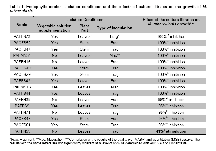
to the experimental control, %; The culture filtrate was considered a strong inhibitor of growth if inhibition was higher than 90% (v/v) and a strong stimulator of growth if the stimulation was higher than 10% (v/v). The assays were carried out in triplicate and analyzed for statistical significance by ANOVA followed by a Fisher's least significant difference (LSD) test. A total of 315 fungal isolates, which represented 85 different morphologies (macro-morphology characterization), were cultured from the leaves and steams of P. aduncum L. One isolate per morphology was selected for the bioprocess. Because 3 isolates could not be purified, a total of 82 isolates were investigated. Some isolates could be identified at the genus level (based on macro- and micro-morphology); for example, members of the Deuteromycetes class including Fusarium, Penicillium and Aspergillus were identified. However, the majority of the fungi did not display reproductive morphologies and were thus difficult to identify. The six P. aduncum L. extracts that were obtained from the stems and leaves using dichloromethane, methanol and water did not influence the growth of M. tuberculosis in either bioassay. Fifteen of the culture filtrates from the endophytic fungi inhibited the growth of M. tuberculosis by more than 90% in the qualitative and quantitative bioassays (Table 1). Of those filtrates, 11 were isolated with culture medium supplemented with the vegetable solution; thirteen were isolated using the fragmentation technique, and six were isolated from the stems and nine from the leaves of P. aduncum L. The Aspergillus PAFFN59 culture filtrate stimulated the growth of M. tuberculosis. After 6 days of incubation, the culture filtrate increased the number of cells of M. tuberculosis by 40%. This strain was isolated from P. aduncum L. leaves using the fragmentation technique and a culture medium without vegetable solution supplementation (Table 1). DiscussionThe plant extracts obtained from the stems and leaves of P. aduncum L. did not contain the compounds with anti-tubercular activity, which have been identified in other species of Piper (P. sanctum, P. aff. pedicellatum and P. sarmentosum) (Mata et al. 2004; Rukachaisirikul et al. 2004a; Rukachaisirikul et al. 2004b; Tuntiwachwuttikul et al. 2006). The possibility that active anti-tubercular substances could be identified in P. aduncum L. tissues was the motivation for this study; however, a parallel study of culture filtrates from the endophytic fungi played an important role in the search for bioactive compounds and became the focus of this study because good results were obtained. The investigation of culture filtrates from 82 endophytic fungi isolated from the stems and leaves of P. aduncum L. identified 15 that inhibited the growth of M. tuberculosis. This result is similar to other studies that evaluated the ability of endophytic fungi to produce metabolites with antimicrobial activity (de Souza et al. 2004). However, the results of 100% growth inhibition in the bioassays can be considered promising because they were obtained with culture filtrates that were not submitted to purification or concentration. In continuing studies, the active metabolites in the culture filtrates should be identified and evaluated as possible antimicrobials. The culture filtrate from Aspergillus PAFFN59 stimulated a 40% increase in the growth of M. tuberculosis in the bioassays. The metabolites present in the culture filtrate of this fungus could be added to the culture media used for classical culturing and in the semi-automated methods used for the identification of M. tuberculosis. This addition would facilitate faster diagnosis of tuberculosis. Using current automated and semi-automated bacteriological methods, it takes 14 to 25 days to diagnose tuberculosis; using classical methods, 25 to 40 days are required (Williams-Bouyer et al. 2000; Piersimoni et al. 2001). This is the first report describing a screen of culture filtrates to identify endophytic fungi that produce metabolites that increase the velocity of M. tuberculosis growth. The knowledge obtained in this work provides insight for future research on the development of a faster method for the diagnosis of tuberculosis.
Copyright 2011 by Universidad Católica de Valparaíso -- Chile The following images related to this document are available:Photo images[ej11045t1.jpg] |
| |||||||||
