 |
| About Bioline | All Journals | Testimonials | Membership | News |
|
||||||
|
||||||
European Journal of General Medicine, Vol. 1, No. 1, Jan-Mar, 2004, pp. 26-27 BRIEF REPORT THE TIME COURSE OF SERUM MALONDIALDEHYDE LEVELS IN BURNED HUMANS Bekir Atik1, Önder Tan1, Haluk Dülger2, Burhan Köseoğlu3, Mehmet Bekerecioğlu4 Yüzüncü Yıl University, Faculty of
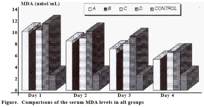
Medicine, Departments of Plastic and Reconstructive Surgery1 Biochemistry2 and Pediatric Surgery3, Gaziantep University, Faculty of Medicine Department of Plastic and Reconstructive Surgery4 Code Number: gm04007 Burn injury is accompanied by complex pathophysiological alterations that exert deleterious effects on various organ systems (1). Inflammatory shock mediators that are implicated in the pathogenesis of burn shock include histamine, serotonin, kinins, oxygen free radicals (OFR), prostaglandins, thromboxane and interleukins (2). There is also experimental evidence documenting superoxide radical involvement in the pathogenesis of burn shock (3). Harmful metabolites (O2, H2O2, O2.) form as the result of lipid peroxidation. OFR are determined by lipid peroxidation products. Malondialdehyde (MDA) is used commonly in determination of lipid peroxidation (4,5). Antioxidant therapy in burn injury has been used to prevent the harmful effect of oxygen free radicals (6). The relationship between burn injury and serum MDA levels is well known, but few studies were conducted about the time course of the oxidant stress. In this study, we aimed to evaluate the time course of serum MDA levels after burning in humans. The study was undertaken in 56 burned patients (mean age 32.5±13.1 years, 1951 years, 32 male) and 20 healthy subjects (mean age 34.0±11 years, 23-54, 12 male, 8 female). The burn injuries were second degree and scalding due to hot liquids. Patients with metabolic disease and sepsis were excluded from the study. Venous blood samples were taken at the first, third, seventh and tenth days of post burn period, and serum MDA levels were measured by tiobarbituric acid method (4) and results were expressed as nanomol (nmol) MDA per milliliter (mL) in serum. The patients were divided into four groups in extent of burn injury (A; 20-25% of body, n:16, B; 26-30 %, n:12, C; 31-35 %, n:18, D; 36-41 %, n:10). The results were evaluated with ANOVA Post Hoc test (Tukey) and Kruskal-Wallis Analysis. In group A, mean serum MDA levels were 11.06 nmol/mL at the first day, 9.21 nmol/mL at the third day, 7.71 nmol/ml at the seventh day, and 5.77 nmol/mL at the tenth day. Mean serum MDA levels were 10.54 , 8.75, 6.87, and 5.83 nmol/mL respectively in group B. In group C, mean serum MDA levels were 11.30, 9.83, 8.75, and 7.12 nmol/mL respectively. Mean serum MDA levels were 12.52, 11.09, 10.06, and 7.18 nmol/mL, respectively in group D (Figure 1). Mean serum MDA level of the control group was 2.95 nmol/mL. Mean serum MDA levels were significantly higher in all groups, for all degrees of burns and for all the time course when compared to controls (for all groups p< 0.0001, except for group B; at 10th day p<0.005) Burn shock results from the interplay of hypovolemia and mediator action and continues as a significant pathophysiologic state, even if hypovolemia is corrected. These physiologic changes can further exacerbate the whole body inflammatory response into vicious cycle of accelerating organ dysfunction (7). In this study it was shown that high MDA levels were seen not only early after the burn injury but even after 10 days. In an animal study, Demling et al. (8) demonstrated that lipid peroxidation was still present at the 5th day of thermal injury. They did not follow up further changes beyond 5 days. Pintaudi et al. (9) showed that high MDA levels persist up o 30 days in humans. Our study, in accordance with these studies, reflects the persistence of oxidative stress beyond the early period of burn injury. Clinical significance of this condition should be investigated and this time course should be considered in design of the studies evaluating the effects of antioxidants in burn patients. REFERENCES
Copyright 2004 - Medical Investigations Society The following images related to this document are available:Photo images[gm04007f2.jpg] [gm04007f1.jpg] |
| |||||||||
