 |
| About Bioline | All Journals | Testimonials | Membership | News |
|
||||||
|
||||||
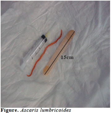
European Journal of General Medicine, Vol. 1, No. 2, 2004, pp. 43-45 CASE REPORT PULMONARY EDEMA ASSOCIATED WITH ASCARIS LUMBRICOIDES IN A PATIENT WITH MILD MITRAL STENOSIS: A CASE REPORT Talantbek Batyraliev1, Beyhan Eryonucu2, Zarema Karben1, Hakan Sengul1, Niyazi Güler2, Orhan Dogru1, Alper Sercelik1 Sani Konukoglu Medical Center , Department of Cardiology1, Yüzüncü Yıl
University, Department of Cardiology2 Code Number: gm04019 Ascaris lumbricoides remains the most common intestinal nematode in the world. Clinical manifestations of ascaris lumbricoides are different in each stage of the infection. We presented an unusual presentation of ascaris lumbricoides. Key words: Pulmonary oedema, mild mitral stenosis, Ascaris lumbricoides INTRODUCTION Ascaris lumbricoides (AL) remain the most common intestinal nematode in the world. Clinical manifestations of AL are different in each stage of the infection. Infection with ascaris appears to be asymptomatic in the vast majority of cases, but may produce serious pulmonary disease or obstruction of biliary or intestinal tract in a small proportion of infected people. We presented an unusual presentation of AL and pulmonary edema (1,2). CASE A 27-year-old woman was admitted to our hospital because of an episode of acute severe dyspnea, nausea and vomiting. Her medical history revealed rheumatic mild mitral stenosis. On admission, the patient was in moderate respiratory distress and slightly agitated. The vital signs were as follows: blood pressure; 125/75mmHg, pulse; 138 beats/min, respiratory rate; 40breaths/min, and temperature; 37.3oC. Auscultation revealed loud S1, a grade 2/6 diastolic murmur. The lung fields had bilateral diffuse crackles. The remainder of the physical examination was normal. Laboratory values were within normal limits. An ECG showed sinus tachycardia. Cardiac enzymes were normal. Chest X ray revealed pulmonary oedema and normal cardiac size. Transthoracic echocardiography was performed to confirm valve function and showed mild mitral stenosis (valve area 1.9cm2) and normal left ventricular function. The patient was treated as acute pulmonary oedema due to mitral stenosis. Treatment of diuretics was initiated. She was placed on oxygen by nasal cannula. Tachycardia was not taken under control by treatment with digoxin and verapamil. A reason for excessive nausea and vomiting was not determined and these semptoms were not responsive to antiemetic drugs. Despite intensive treatment, clinical improvement was not occured. Fortunately, at the 3rd day, the patient was expelled ascaris lumbricoides (AL) (Figure). Once the AL was removed the patient’s respiratory condition dramatically improved. The patient was started on a 3-day course of mebendazole and discharged 4 days later in good general condition. DISCUSSION Severe pulmonary edema due to mild mitral stenosis is an unexpected finding. In this case, we suspected pulmonary edema due to mitral stenosis because of the medical history of the patient at admission. Expiration of AL helped diagnosis and treatment of this patient. Dramatic and rapid clinical improvement after expelling of AL with vomitting indicated that the reasons of pulmonary edema were excessive tachycardia, shortened diastolic filling period and elevated left atrial and pulmonary venous pressure associated with nausea and vomiting. To our knowledge pulmonary edema associated with AL has not been reported. Ascariasis is caused by the parasite AL, the largest intestinal nematode found in humans. Ascariasis remains the most common intestinal nematode in the world. It is widely distributed in tropical and subtropical regions where there is insufficient sanitation, hygiene, and education regarding these parasites. Ascariasis generally occurs through hand-to-mouth ingestion of agricultural products or food contaminated with parasite eggs. Rarely, transmission can occur via inhalation of eggs or swallowing contaminated respiratory secretions. The life cycle of AL is complex and begins with the ingestion of eggs. The larvae penetrate the intestinal wall and enter surrounding capillaries where they migrate through the lungs 1–2 weeks after the initial ingestion. In the lungs, the larvae again penetrate the capillary walls, entering the alveolar space where they ascend the tracheobronchial tree to the epiglottis, where they are eventually swallowed. The larvae reach the small intestines, most commonly the jejunum, where they mature into adult worms. They live from 10 to 24 months and are produce up to approximately 240,000 eggs per day 2–3 months after the initial infection. Adult forms may reach up to 40 cm in length and live for two years (1). Several clinical manifestations of this infection are reported. Typically, complications occur as a result of a large obstructing mass of worms in the intestines, the induction of stones by a single worm in the hepatobiliary tree, or an inflammatory reaction with the migration of adult worms and larvae through tissue. During the lung phase of larval migration, about 9 to 12 days after egg ingestion, patients may develop an irritating nonproductive cough and burning substernal discomfort. Fever is common. Eosinophilia develops during this symptomatic phase. Larval pulmonary migration is generally asymptomatic. However, symptomatic pulmonary disease may occur with fever, cough, chest pain, hemoptysis, dyspnea, and wheezing due to (1) Loffler’s syndrome, (2) the effects of larval tissue migration, (3) airway reactivity or bronchospasm, (4) infectious bacterial complications from parasitic migration and associated aspiration, and rarely (5) chronic eosinophilic pneumonia, transdiaphragmatic penetration, or symptoms of upper airway obstruction. Clinical evaluation shows pulmonary opacities on chest radiograph, peripheral blood eosinophilia, and larvae in respiratory or gastric secretions (1-3). Loeffler’s syndrome is another clinical manifestation of AL infection. It is most commonly seen in children and presents as cough; dyspnea; wheezing; sudden-onset fever; substernal chest discomfort; and less commonly, hemoptysis. Symptoms develop 9–12 days after ingestion of the eggs and last 2 to 3 weeks. The hallmark of Loeffler’s syndrome is fleeting pulmonary infiltrates on chest X-ray, often accompanied by peripheral eosinophilia (3). The most important tools for the diagnosis of AL infection are history, chest X-ray, and ultrasonography. Diagnosis is by microscopic detection of eggs in fecal samples. Occasionally, patients present after passing an adult worm in the stool or through the mouth or nose. During the early transpulmonary migratory phase, larvae can be found in sputum or gastric aspirates. AL may enter into nasogastric and endotracheal tubes and block their lumen (4-6). The classic treatment choices are mebendazole orally twice a day for 3 days, albendazole orally as a single dose, or pyrantel pamoate orally as a single dose not to exceed 1 g. A stool ova and parasites test should be repeated 2 weeks after treatment to ensure adequate eradication of the helminth because these medications typically do not have an effect on the larvae. Pulmonary ascariasis is typically a self-limited disease requiring only supportive therapy. However, if there is a superimposed bacterial infection, appropriate antimicrobial therapy is warranted. Antibiotics also may be indicated in the initial management of a patient with such infiltrates until the diagnosis is firmly established (1,2). REFERENCES
Copyright 2004 - Medical Investigations Society The following images related to this document are available:Photo images[gm04019f1.jpg] |
| |||||||||
