 |
| About Bioline | All Journals | Testimonials | Membership | News |
|
||||||
|
||||||
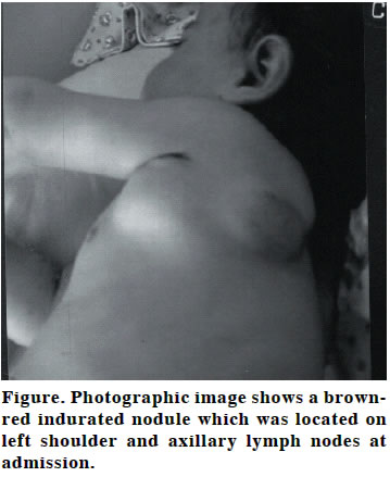
European Journal of General Medicine, Vol. 1, No. 2, 2004, pp. 46-47 CASE REPORT ACUTE LYMPHOBLASTIC LEUKEMIA PRESENTING WITH CUTANEOUS INVOLVEMENT Ali Bay, Ahmet Faik Öner, Oğuz Tuncer, Bülent Ataş, Sevil Yuca, Mehmet Erol Yuzuncu Yıl University, Faculty of Medicine, Department of Pediatrics Code Number: gm04020 Primary cutaneous involvement in B-cell lymphoblastic leukemia is rare in childhood. We present a case of 2-year-old girl admitted to our hospital because of left scapular skin lesion and left axillary mass. She was looked pale and weak. Left axillary lymph node pachage of 5 cm diameter and left scapular skin lesion was revealed by physical examination. Complete blood count revealed 124.000/mm3 white blood cell count. Peripheral blood smear showed 99% lymphoblasts. Bone marrow aspiration revealed 95% blastic cells with immunophenotype and morphological characteristics of pre-B type acute lymphoblastic leukemia with L1 subtype. ALL St Jude Total XIII remission induction treatment protocol was started. Skin lesion disappeared after 15th day of the cytotoxic therapy. On the follow up, she was on remission and continued to maintenance chemotheraphy for 6 months. We would like to highlight that a small growing cutaneous lesion could be the presenting form of acute lymphoblastic leukemia. Key words: Lymphoblastic leukemia, cutaneous involvement INTRODUCTION Skin involvement can be the initial symptom of hematologic malignancies in children (1). Cutaneous involvement in children with acute lymphoblastic leukemia (ALL) is very rare condition compared to acute myeloblastic leukemia (AML). Cutaneous infiltrates in children with acute monocytic leukemia is well-known (2). Scant information is available concerning the occurrence and natural history of skin involvement in children with B-cell ALL. We reported a case of ALL presented with left scapular skin lesion. CASE A 2-year-old girl admitted to our hospital because of left scapular skin lesion and left axillary mass. She complaint fatique, bone pain and anorexia for 4 months and progressing scapular and axiller mass which the parents recognized 10 days before. An operation was planned for surgical resection by plastic surgery team. High leukocyte count was found in rutin preoperative blood analysis and the patient referred to hematology clinic. She was looked pale and weak. Left axillary lymph node pachage of 5 cm diameter and left scapular skin lesion was revealed by physical examination. The skin lesion was asymptomatic brown- red indurated nodule of 5cm diameter without excoriation or ulceration (Figure). She had an enlarged liver and spleen both palpable 3cm below the costal margin at the mid clavicular line. The remainder of the physical examination was unremarkable. Results of the complete blood count were as follows; Hemoglobin: 7.4g/dL, white blood cell count: 124.000/mm3, and platelet count 40.000 /mm3. Peripheral blood smear showed 99% L1 type lymphoblasts. ALT was 104 IU/L, AST was 258 IU/L, lactic dehydrogenase was 15200 IU/L, uric acid was 9mg/dl with normal renal function tests. Chest roentgenogram and bone survey were normal. Abdominal ultrasonography was normal with the exception of hepatosplenomegaly. Examination of bone marrow aspirate showed 95% blast cells that were characteristic of ALL, with L1- type morphology. We found CD10 and CD19 values were positive whereas CD5, CD7, CD13, and CD20 values were negative on flowcytometry. The leukemic character of skin infiltration was confirmed by skin needle aspiration. With these findings, morphologically L1 and immunologically preB type ALL was diagnosed. ALL St Jude Total XIII remission induction treatment protocol was started. Skin lesion disappeared after 15th day of the cytotoxic therapy. On the follow up she was on remission and continued to maintenance chemotheraphy for 6 months. No recurrence of the skin lesion was seen during this period. DISCUSSION Various cutaneous lesions can be observed in patients with hematologic malignancies. These include specific cutaneous lesions resulting from infiltration of the skin by the malignant cells, characteristic diseases such as pyoderma gangrenosum and Sweet syndrome, cutaneous signs of infection or hemorrhage resulting from the bone marrow dysfunction induced by the malignant process or chemotherapy. The frequency of leukemic infiltration of the skin is variable according to the type of leukemia. Cutaneous involvement is a common finding in AML with monocytic differentiation while it is a rare event in ALL (3). Dunn et al. (4) reported 2 patients with leukemic infiltration of the skin among 40 children with ALL. In another study, 15 patients with initial leukemic infiltration of the skin among 1359 children with ALL was reported (5). There was no agreement on association between skin lesions and prognosis (5-7). Cutaneous involvement can be observed not only among high risk ALL patients but can be also observed among low risk patients. Millot et al. (3) reported 9 of the 15 children with initial leukemic infiltration of the skin have unfavorable prognostic factors like high leukocyte count, hepatosplenomegaly and age under 12 months. The skin lesions of these patients can be regarded as a dissemination of aggressive leukemia to the skin. Su et al. (6) reported that; appearance of specific skin lesions in leukemia is usually associated with a very poor prognosis. In a previous report of 25 cases of ALL in infants, skin infiltration was associated more closely with the patient’s age (early period of infancy) than with karyotype abnormalities (8). In our patient, high leukocyte count was thought to be as a poor prognostic risk factor. In the patient, skin lesion was disappeared within 15 days after the cytotoxic therapy and complete remission was obtained after induction theraphy. We would like to highlight that a small growing cutaneous lesion could be the presenting manifestation acute lymphoblastic leukemia. REFERENCES
Copyright 2004 - Medical Investigations Society The following images related to this document are available:Photo images[gm04020f1.jpg] |
| |||||||||
