 |
| About Bioline | All Journals | Testimonials | Membership | News |
|
||||||
|
||||||
European Journal of General Medicine, Vol. 1, No. 3, 2004, pp. 37-42 ORIGINAL ARTICLE PROTECTIVE EFFECTS OF QUERCETIN, A FLAVONOID ANTIOXIDANT, IN ABSOLUTE ETHANOL-INDUCED ACUT GASTRIC ULCER Ömer Coşkun1, Mehmet Kanter1, Ferah Armutçu2, Kurtuluş Çetin1, Betül Kaybolmaz1, Ömer Yazgan1. Karaelmas University, Faculty of Medicine, Departments of Medical Histology-Embryology1 and
Biochemistry2 Code Number: gm04030 Free radicals have been reported to be responsible for many ailments including gastroduodenal ulcers. Recent studies demonstrated that quercetin (QE) has an antioxidant effects on injuries caused by various toxic agents in different experimental models. In our study, we examined anti-ulcerogen and antioxidant effects of quercetin on ethanol-induced gastric lesions in rats. QE was administered intraperitoneally (i.p.) 50 mg/kg. It was found that pretreatment with QE significantly reduced ethanol-induced gastric damage and malondialdehyde levels, and significantly increased antioxidant enzyme activities. We conclude that QE clearly has antioxidant properties and that the protective effect of QE against ethanol-induced gastric mucosal lesion, at least in part, depends upon the reduction in the lipid peroxidation and an increase in the activity of antioxidant enzymes. Key words: Quercetin, ethanol, antioxidant enzymes, gastric damage, oxidative stress, rat. INTRODUCTION Ethanol manifests its harmful effects either through direct generation of reactive metabolites, including free radical species that react with most of the cell components, changing their structures and functions, or by contributing to other mechanisms that finally promote enhanced oxidative damage (1,2). Ethanol-induced gastric mucosal injury is associated with extensive damage to mucosal capillaries and increased vascular permeability (3,4). Mucosal capillary necrosis, vascular congestion and thrombosis in the subepithelial microvasculature accompany disruption of the gastric mucosal barrier. In addition to the direct injurious effects of ethanol on gastric mucosa, other factors are also thought to be involved in the pathogenesis of injury (5). Flavonoids are a group of naturally occurring compounds widely distributed as secondary metabolites in the plant kingdom. They have been recognized for having interesting clinical properties, such as anti-inflammatory, antiallergic, antiviral, antibacterial, and antitumoral activities (6). One of these flavonoids, quercetin (3,5,7,3’,4’-pentahydroxyflavone), prevents oxidant injury and cell death by several mechanisms, such as scavenging oxygen radicals (7,8) protecting against lipid peroxidation (9) and chelating metal ions (10). Quercetin, a flavonoid found in many plants, is widely distributed in edible fruits and vegetables. When quercetin was administered orally, it was poorly absorbed from the digestive tract and did not have a great influence on the organs (11,12). Quercetin is the major flavonoid in the human diet and its daily intake with foods is estimated to be 50–500 mg (13). Its preventive roles in experimental models of various agent-induced damage, including ethanol, gastric ulcers have been reported (14-16). This study aimed to examine antiulcerogen and antioxidant effects of quercetin on ethanol-induced gastric lesions in rats. For this purpose, we studied the effects of quercetin antioxidant activity, and changes in lipid peroxidation which were determined by measuring MDA level. MATERIALS AND METHODS Animals Table 1. Erythrocyte MDA (µmol/g Hb), serum NO (µmol/l) and gastric tissue MDA (µmol/g protein), SOD (U/mg protein), GSH-Px (U/mg protein) and CAT (k/mg protein) levels of all groups. A (control), B (ethanol) and C (ethanol+quercetin 50 mg) groups. Values are expressed as mean ± SD, and n = 10 for all groups.
a P<0.05 when compared with group A, b P<0.05 when compared with group A and B, c P<0.05 when compared with group B All animals received human care according to the criteria outlined in the “Guide for the Care and Use of Laboratory Animals”prepared by the National Academy of Sciences and published by the National Institutes of Health. Treatment of rats One hour after the administration of ethanol, animals were sacrificed by cervical dislocation and the stomach of each was removed and opened along the greater curvature and washed in physiological saline solution. For the measurement of the gross gastric mucosal lesions, freshly excised stomach was laid flat and the mucosal lesions were traced on clear acetate paper. Gross mucosal lesions were recognized as haemorrhage or linear breaks (erosions) with damage to the mucosal surface. The area of stomach and gross lesions were calculated by image analysis system. The system used is composed of a PC, hardware and software (Image-Pro Plus 5.0-Media Cybernetics, USA) for image acquisition and analysis, Spot Insight QE (Diagnostic Instruments, USA) camera and optical microscope. The method requires preliminary software procedures of spatial calibration (micron scale) and setting of color segmentation for quantitative color analysis. Table 2. Comparison of the area of the gastric lesion in A (control), B (ethanol) and C (ethanol+quercetin 50 mg) groups. Kruskal-Wallis test was used for statistical analysis. Values are expressed as mean ± SD, and n = 10 animals for all groups.
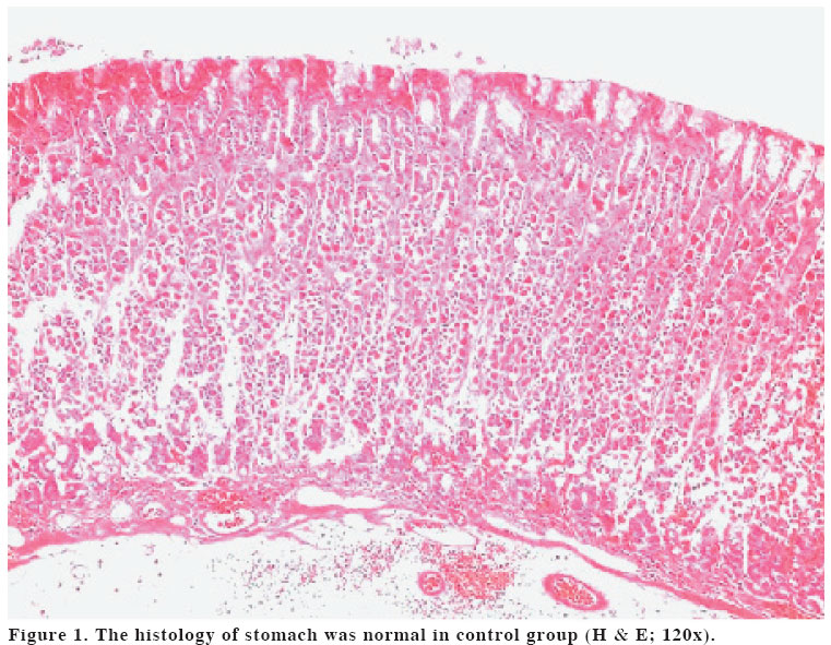
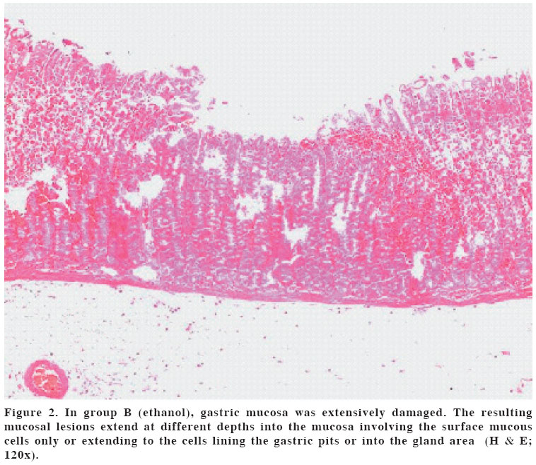
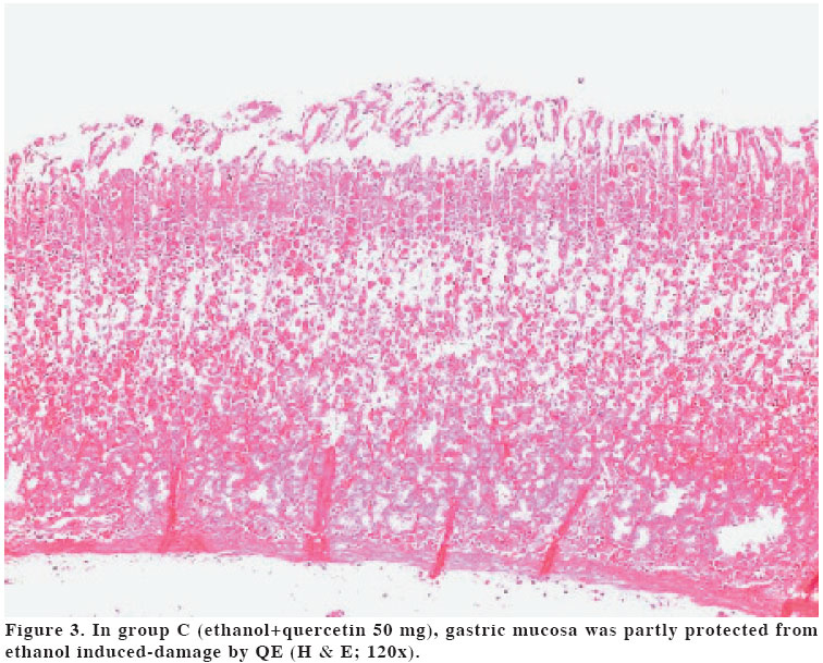
a P<0.01 when compared with group A and B Biochemical analysis Histopathological procedures Statistical analysis RESULTS Biochemical findings Histological findings In group B, gastric mucosa was extensively damaged. The resulting mucosal lesions could vary in that they could extend at different depths into the mucosa involving the surface mucous cells only or extending to the cells lining the gastric pits or into the gland area (Figure 2). In group C, gastric mucosa was partly protected from ethanol induced-damage by QE (Figure 3). DISCUSSION Oxygen derived free radicals have been implicated in the pathogenesis of a wide variety of clinical disorders and gastric damage is caused by physical, chemical and psychological factors that leads to gastric ulceration in human and experimental animals (23). Most of the available drugs are thought to act on the offensive factors which neutralize acid secretion like antacids, H2 receptor blockers like ranitidine, famotidine, anticholinergics like pirenzepin, telezipine, proton pump blockers like omeprazole, lansoprazole, etc. which interfere with acid secretion (24). The present study was designed to demonstrate gastric ulcer protective activity of quercetin in rats. Quercetin is a phenolic compound widely distributed in the plant kingdom. It is found in frequently consumed foods, including apples, berries, onions, tea and brassica vegetables. Quercetin is reported to have many beneficial effects on human health, including cardiovascular protection, anticancer activity, antiulcer effects, antiallergic activity, cataract prevention, antiviral activity and anti-inflammatory effects (25-27). In the present study, quercetin significantly decreased the concentration of malondialdehyde, an indicator of lipid peroxidation, in the stomach homogenates. Among the numerous pathological events that are caused by an imbalance between oxidative damage and antioxidant defense systems, lipid peroxidation is a wellknown example of oxidative damage that affects cell membranes, lipoproteins, and other lipid-containing structures under conditions of oxidative stress. Therefore, the fact that quercetin decreased lipid peroxidation might mean that quercetin has an antioxidant effect. Quercetin has effective antiulcer activity against ethanol-induced ulcerogenesis and it can be used for therapy of ulcerogenesis and gastric mucosal injury. Quercetin protects and prevents changes in biochemical parameters and morphologic changes observed after ethanol consumption. In our study, erythrocyte and tissue MDA and serum NO concentration was significantly increased in ethanol group with reduction in antioxidant enzyme activities of SOD, GSHPx and CAT. QE treatment decreased the elevated MDA and NO and also increased the reduced antioxidant enzyme activities. Namely, our results indicate that the preventive effects of QE may be due to inhibition of lipid peroxidation by its antioxidant nature. As conclusion, QE shows protective effects in ethanol-induced gastric ulcer, possibly by decreasing oxidative stress and increasing antioxidant enzyme activity. But to elucidate the exact mechanism of this modulatory effect, and to examine its potential therapeutic effects further studies are essential. REFERENCES
Copyright 2004 - Medical Investigations Society The following images related to this document are available:Photo images[gm04030f3.jpg] [gm04030f2.jpg] [gm04030f1.jpg] [gm04030t2.jpg] [gm04030t1.jpg] | ||||||||||||||||||||||||||||||||||||||
| |||||||||
