 |
| About Bioline | All Journals | Testimonials | Membership | News |
|
||||||
|
||||||
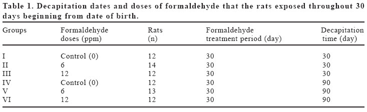
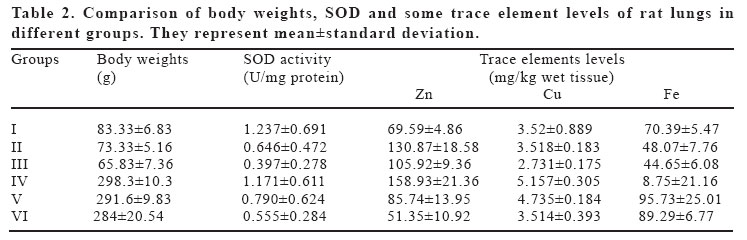
European Journal of General Medicine, Vol. 2, No. 2, 2005, pp. 62-68 THE CHANGES OF ZINC, COPPER, AND IRON LEVELS IN LUNG TISSUE AFTER FORMALDEHYDE INHALATION DURING THE EARLY POSTNATAL PERIOD OF RATS Ahmet Songur1, İlter Kuş2, Şemsettin Şahin3, Sadık Söğüt4, Oğuz Aslan Özen1 Mehmet Yaman5, Mustafa Sarsılmaz2 Afyon Kocapete University, Faculty of Medicine, Department of Anatomy, Afyon1, Fırat University, Faculty of Medicine, Department of Anatomy, Elazığ2, Gaziosmanpaşa University, Faculty of Medicine, Department of Biochemistry, Tokat3, Mustafa Kemal University, Faculty of Medicine, Department of Biochemistry, Hatay4, Firat University, Faculty of Science and Arts, Department of Chemistry, Elazığ5, Turkey. Correspondence: Dr. Ahmet SONGUR Afyon Kocatepe Üniversitesi,
Tıp Fakültesi Anatomi A.D. Afyon, TURKEY Code Number: gm05013 Aim: In this study, effects of inhaled formaldehyde (FA)
gas, during early postnatal period on the levels of zinc, copper and iron elements
and activity of total superoxide dismutase (t-SOD) enzyme in lung tissue and
also the reversibility of effects of formaldehyde were examined. Key words: Formaldehyde gas, lung, t-SOD, zinc, copper, iron INTRODUCTION Formaldehyde (FA) is a colorless gas with pungent smell and is accepted as toxic over certain concentrations. Harmful effects in the room temperature was due to its easy evaporation (1). FA is present in nature, in cigarette smoke, in domestic air, in the polluted atmosphere of cities (2,3) and also, it is widely used in industrial and medical settings and employees may be highly exposed to it in these settings. Especially anatomists and medical students having dissection lectures are most frequently subjects to exposed FA gas. It is known that FA induces a variety of toxic effect on biological system, such as respiratory tract and nervous system irritations, genotoxicity, cytotoxicity and carcinogenicity (2,3). Inhaled FA primarily effects the airways and has negative effects on respiratory system, and these effects depends on its concentration in the air. It is highly irritant to the mucous membranes and its effect on the upper respiratory tract is well documented (4). In the workplace, exposure to FA has been implicated in occupational asthma. Domestic exposure to FA has been associated with lower respiratory tract symptoms and disease in both children and adults (5). Specific mechanisms to account for these findings have not yet been identified; however, it is possible that formaldehyde may have a direct toxic effect on the respiratory epithelium, inducing inflammatory reactions. Inflammatory changes are observed in the upper airways after acute low-level exposure to formaldehyde, and damage to the lower airways is reported after exposure to high levels (5 to 30 ppm) (5). Superoxide dismutase (SOD) is one of the most important enzymes that function as cellular antioxidants. It is present in cell cytoplasm and in mitochondria in order to maintain a low concentration of superoxide anion which is the major radical of oxygen (6). Zinc, copper and iron elements have important role in tissue as co-factors of some enzymes. Zinc and copper is functioning as cofactors of cytoplasmic Cu-Zn superoxide dismutase enzyme. If the concentrations of these trace elements are reduced significantly, SOD can not detoxify the harmful oxygen species (7,8). Additionally, increase or decrease of these trace elements in the tissues seriously damage and cause malfunctions. They are the signs explaining the condition of the tissue and these overtake important tasks (8,9). It has been observed that the increase or decrease of these elements’amount in the lung tissue causes various damages. But the effect of inhaled FA throughout developmental process on Zn, Cu and Fe levels in lung tissue is interesting. Therefore, the aim of this study was to investigate i) the effect of inhaled FA gas during early postnatal period on the changes of zinc, copper and iron elements ii) the effect of FA gas on activity of SOD enzyme in the lung tissue of rats in developmental process iii) to determine, if any, whether the changes is reversible or not. MATERIAL AND METHODS Three vitreous quadrangular chambers (20x50x100 cm) were prepared. Two holes were opened on each chamber for inflow and outflow of air. By the help of air pumps the circulation of the air in the chambers is fixed 10 L/min. The FA gas was generated by heating paraformaldehyde (Merck KGaA, Darmstadt, Germany) at 70–90 0C according to a method described by Chang et al. (10). It was pumped from production area to chambers on desired levels and time. Healthy and mature male and female albino Wistar rats were obtained from Firat University Biomedical Research Unit of the Institute of Health Sciences, were mated. The groups were arranged with pups, born at the same time, and put in the chambers after birth. The FA gas was given into chambers at concentrations 0 (control), 6 or 12 ppm for 30 days (6 hours/day; 5 days/week). Its concentrations were regularly monitored with the formaldehyde monitor (Enviromental Sensors Co., Boca Raton FL 33431 USA). After the treatment the rats were divided in six groups for decapitation at 30th and 90th days (Table 1). Pups were kept in chambers for two weeks continuously with their mothers. After two weeks they were kept in plastic cages and transferred into the chambers only during the treatment. Vitreous chambers and plastic cages were cleaned regularly. Water (tap water) and food (Institutes’stock diet included 24 mg Zn, 16 mg Fe and 2 mg Cu in Per 1 gr) were replaced every day. The rats were checked daily and body weights were recorded every week and just before the decapitation. The temperature of the vitreous chambers were fixed to 25 ± 2 0C, the humidity level was 40-50% and light was 12 h day / 12 h night cycle. Food and water were provided ad libitum during the FA treatment and rest of the time. At the decapitation day, 6 male rats from each group were killed by decapitation under ether anesthesia and the lungs were removed immediately and weighed and were fixed in 10% formaldehyde solution. For trace elements analysis; an ATI UNICAM 929 Model atomic absorption spectrophotometer (AAS) equipped with ATI UNICAM hollow cathode lamp was used for the determinations. Unless stated otherwise; all chemicals used were of high-purity reagent grade. In all analytical work, double distilled water was used. All glass materials were kept stored with 1 mol.L-1 nitric acid when not in use. In the digestion and extraction procedures, concentrated nitric acid (65%, Merck) and hydrogen peroxide (35%, Merck) were used. A stock solution of Zn, Cu and Fe (1000 mg L-1) was prepared by dissolving Zn(NO3)2, Cu(NO3)2 and Fe(NO3)2 in 1.0 mol L-1 nitric acid. For the accuracy check of the metal analysis, standard addition and calibration methods were applied. The slopes of calibration graph obtained with the matrix components added to standard solutions are found to be similar to those obtained with standard addition methods for each three metals. Therefore, applied analyze method is accurate and reliable. In order to prevent trace metals contamination; the beakers were cleaned before taking lung tissue samples. The beakers were washed with chromic acid, rinsed with distilled water. Then, 1.0 cc HNO3/H2O2 mixture (2/1) was placed into the beakers and evaporated until a 0.1-0.2 cc left over residue and the bakers were washed with distilled water again. 0.1-0.2 g of tissue samples was placed into the beakers and 0.5 cc concentrated HNO3/H2O2 mixture (2/1) was added. Then, each beaker was carefully shaken and boiled until 0.1-0.2 cc residue was left. Clear residue was completed to 1.0 cc with 1.0 mol.L-1 HNO3. After necessary dilutions, Zn, Cu and Fe concentrations of solution were determined by AAS. Blank digests were carried out by the same way. For biochemical analysis, after weighing the lungs, tissues were homogenized in a four volumes of ice-cold Tris-HCl buffer (50 millimolar, pH 7.4) containing 0.50ml/L Triton X-100 with a homogenizator (IKA Ultra–Turrax T 25 Basic, Germany) for 2 minutes at 13000 rpm. The homogenate was then centrifuged at 5000 x g for 20 minutes to remove debris. Clear supernatant was taken and for a further extraction procedure, it was extracted in ethanol/chloroform mixture (5/3, v/v). After second centrifugation at 5000 x g for 20 min, the clear upper layer (the ethanol phase) was taken and used in SOD activity protein amount determination. Total SOD (EC 1.15.1.1) activity was determined according to the method of Sun et al. (11) with a modification by Durak et al. (12). The principle of the method is based on the inhibition of nitroblue tetrazolium (NBT) reduction by the xanthine-xanthine oxidase system as a superoxide generator. One unit of SOD was defined as the enzyme activity causing 50% inhibition in the NBT reduction rate. SOD activity was expressed as units per mg tissue protein (specific activity). Protein measurements were made in tissue homogenate according to the method of Lowry et al. (13). Statistical analysis was performed on a personal computer using SPSS for Windows software. Kruskal-Wallis H Test was used to compare continuos variables between the each groups. Intergroup comparisons were performed using Mann-Whitney U Test. p<0.05 was considered statistically significant. RESULTS There was no difference on the mortality rates between the all groups (1 or 2 rat in the each group). In the rats inhaled FA for 30 days period (Group II and III); sneezing, dispnea, polipnea, increase in nose cleaning, excessive licking and blinking of eyes, and nasal mucosa bleeding were observed throughout the treatment. Food and water consumption reduced in these rats than control rats (Group I) relatively. These findings reported above were decreased after the cessation of FA inhalation and diminished throughout the rest 90 days of the treatment (Group V and VI). The body weights of rats in Group II and III were significantly decreased compared to Group I rats. However, Group III body weights were less than Group II, these differences did not reach statistically significance (p=0.07). In Group V and VI rats, there was no statistically significant difference compared to Group IV rats (Table 2). Tissue t-SOD activity was reduced in Group II rats compared to control group, but it was not statistically significant. t-SOD activity was reduced significantly in Group III rats (p<0.007). In the 90th day groups, SOD activity was found to be reduced both two FA dose groups but these findings were not statistically significant (Table 2). In a general view, Cu and Fe concentrations of lung tissue were decreased after FA inhalation in both 30th and 60th days groups. In 90th day treatment groups, Cu levels were decreased gradually depending on FA inhalation dose, that is, there were significant decreases in Cu level in Group V and Group VI compared to control group. It was also found a significant difference between 6 and 12 ppm dose groups. As seen in Table 2, Fe concentrations in 30th day rats were found to be decreased in 6 ppm and 12 ppm groups when compared with control group. Interestingly, Zn concentrations in 30th and 90th day experiment groups were contrary when compared each other. While there was an increase in 30th day FA inhaling rats, there was a diminish in 90th day groups. All the changes in the element concentrations of the groups were statistically significant (p values changes between 0.026 and 0.002) (Table 2). When the element values were compared according to the decapitation time, it was obviously seen that all the element concentrations were higher in the control group of three-month experiments that those of one-month experiment group. This should be result from the age of the animals. DISCUSSION FA is an avid linker of other molecules, including DNA, proteins and unsaturated fatty acids and may cause inflammatory reactions, cancer and other deleterious health effects (14,15). The majority of inhaled FA is near absolutely absorbed in nasal mucosa, trachea and bronchi, and it primarily affects the eyes and upper respiratory tracts. The histopathologic changes in the nasal cavity due to FA inhalation are well documented. Inflammatory cellular changes in the upper respiratory tract of both humans and animals, and nasal squamous cell carcinoma in animals have been observed to be associated with FA inhalation (14,16). However, formaldehyde-induced lesions of the lower respiratory tract are different in species. For example lower airway inflammation has not been observed in mice or guinea pigs after formaldehyde exposure, but has been detected in the trachea and major bronchi of monkeys. In humans, lower airway and pulmonary effects are observed after exposure to formaldehyde at levels greater than 5 ppm (17). But also, chronic low level formaldehyde exposure may cause inflammatory response in the lower airway. Franklin and colleagues (5) demonstrated that exposure to formaldehyde, at domestic environment levels, is associated with raised exhaled nitric oxide levels, a marker of lower airway inflammation, in healthy children. Playing an important role in a number of biological processes, trace elements are an essential part for human being. Trace elements have been extensively studied in recent years to assess the relationship in the etiology of cancer or other diseases. Evidences in recent years demonstrate the ability of several carcinogenic metals to interact with the genetic components of the cells. Most metals that are carcinogenic in humans and animals such as Ni, Cr, Cd and Pb exhibit low to moderate mutagenic activity in many assay systems (18). Although oxidative mechanisms have been implicated in metal carcinogenesis, cells and tissues are generally well protected from excessive oxidation products (19,20). A comparison of trace element levels in normal and injured tissues has demonstrated the existence of statistically meaningful differences between trace element concentrations. In this study, the concentrations of copper, zinc, and iron were determined in the tissues exposed to FA comparing with the controls. The levels of all three elements analyzed in the lung tissue were considerably lower than in the control group. Iron participates in a number of biochemical pathways, provides a prosthetic group for various enzymes particularly antioxidant enzyme catalase, and important in virtually all aspects of life from blood oxygenation, oxidative phosphorylation process to xenobiotic mechanism. Iron is even more critical to the growth of tumor cells when its control is disregulated (21,22). In addition, Fe incorporated heme group is a cofactor for peroxisomal antioxidant enzyme catalase. This enzyme contains a heme group in its active site responsible for its catalytic activity (23). Decreased Fe content in FA-exposed lung tissue may have a negative influence on CAT activity and result in excessive H2O2 accumulation. According to our results, the fact that Fe levels were decreased in lung tissues (especially in 30th day group) may suggest the decreased activity of catalase. We can conclude from these observations that increasing H2O2 production by decreasing CAT activity can be a favorable event as long as the cell does not have to cope with an oxidant stress. Copper and zinc concentrations in lung tissues were found to be decreased depending on the concentration of FA compared to control tissues in the present study. Copper is an essential cofactor for several enzymes such as cytochrome C oxidase, lysyl oxidase etc. Likewise, zinc is a cofactor for many enzymes and a component of several metalloproteins including the important class of regulatory zinc finger proteins. In contrast to copper, zinc is not highly toxic to mammalian cells except at extreme levels (24). Zinc is involved in many biochemical functions. The effect of zinc on protein synthesis may be attributable to its vital role in nucleic acid metabolism. The activities of many zinc-dependent enzymes have been shown to be affected adversely in zinc-deficient tissues. Zinc atoms in some of the enzyme molecules participate in catalysis and also appear to be essential for maintenance of structure of apoenzymes. Zinc also plays a role in stabilization of biomembrane structure and polynucleotide confirmation (25). On the other hand; dietary zinc has been documented to play a role in protecting against hyperoxia-induced lung damage (26). The catalytic activity of free radical scavenging enzyme SOD depends on the presence of a prosthetic group containing copper, zinc stabilizes the apoenzyme in the native configuration. SOD catalyzes the dismutation of superoxide radical (O2.-) into hydrogen peroxide (H2O2). The decreased copper and zinc levels in lung tissue in comparison with unexposed lung tissue may show the decreased capability of the enzyme against oxidative stress. The effect of inhaled formaldehyde on the zinc levels may show the functional and / or structural harmful effects of formaldehyde on lung tissue. Protection of cellular components from damage by reactive oxygen species (ROS) can be accomplished through enzymatic and nonenzymatic defense mechanisms. Intracellular distribution of these systems is very important in the decomposition of toxic reactive intermediates because nucleic acids, proteins, and some enzymes in the cell are found to be attacked by ROS. Active ROS may also damage specific genes that control the growth and differentiation during promotion phase and stimulate a more rapid growth and malignancy of cells (6). So, it can be suggested that carcinogenesis might be induced by ROS. Regarding SOD enzyme activities in cancer tissue, decreased activity was established in most of the publications (27,28). It is also possible to find some reports indicating increased activities of free radical scavenging enzymes in the cancerous cells and tissues (29,30). Low SOD activity obtained in this study supported the hypothesis that enzymatic antioxidant defense mechanisms were impaired in FA-exposed lung tissue, although this was not a universal characteristic of neoplastic changes of the tissue. The question here is may changes in free radical metabolism play a part in the carcinogenic process? It seems quite possible that disordered metabolism in FA-exposed cells and tissues may also lead to inhibition or repression of the synthesis of ROS-metabolizing enzyme proteins. The clinical findings occurred during the treatment in this study were mostly due to irritation of respiratory system and eyes. Our previous studies showed similar findings (8,9). Moreover, the researches conducted by Wilmer et al. (31) and Woutersen et al. (32) confirm these findings. Decrease in clinical findings after the treatment and diminish in Group V and VI rats suggested that the irritation due to FA gas is not permanent. The body weight loss and decrease in food and water consumption in Group II and III rats were an interesting point. In our previous studies, it was showed that inhaling FA caused decrease in body weight gain (8). Also, Martin (33) and Saillenfeilt et al. (34) found that inhaling of FA (10-40 ppm) during the gestation term caused decrease in maternal food consumption, decrease in body weight gain and pups with low born weight. The sign of recovery in Group V and VI rats showed that the rats’bodies can repair themselves after stopping of the FA gas. We think that exposure to formaldehyde may cause growth retardation and alter the trace element levels of lung tissue including copper, zinc and iron, and induce further oxidative damage on lung tissue. ACKNOWLEDGEMENT: The authors thank Dr. Ömer Akyol for critical revision. REFERENCES
Copyright 2005 - Medical Investigations Society |
