 |
| About Bioline | All Journals | Testimonials | Membership | News |
|
||||||
|
||||||
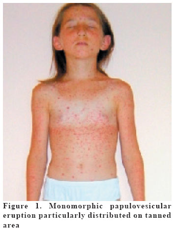
European Journal of General Medicine, Vol. 2, No. 2, 2005, pp. 89-90 PHOTODISTRIBUTED VARICELLA Şemsettin Karaca1, Mustafa Kulaç1, Ömer Doğru2, Zafer Çetinkaya3 Afyon Kocatepe University, Medicine Faculty, Departments of Dermatology1, Pediatric Diseases2 and Microbiology3, Afyon, Turkey Correspondence: Semsettin KARACA, MD Afyon Kocatepe
Universitesi, Ahmet Necdet Sezer Uygulama ve Arastırma Hastanesi
Dermatoloji Klinigi, Afyon/Turkey Code Number: gm05019 Varicella is a common viral disease of childhood with typical clinical presentation. Some of viral exanthemas can be aggravated by sun exposure, trauma and inflammation. Photodistribution of varicella is rare. We report a case of 11 year-old girl who presented with varicella particularly distributed on sun exposed area. Key words: Photolocalized, photodistributed, varicella INTRODUCTION Varicella, one of the viral exanthemas of childhood, is known to have a predilection for sun exposure (1), traumatized (2) and inflamed areas (3). The phenomenon of photosensitivity associated with infectious diseases has been recognized for many years. Photodistributed varicella infection is rare and the typical polymorphism of varicella lesions may not be present. Unfamiliarity with this phenomenon may lead to misdiagnosis as a specific photodermatosis of non-infectious etiology (4). A case of varicella that was aggravated by ultraviolet light exposure is presented. CASE A previously healthy 11 year-old girl was admitted to the dermatology out-patient department with a 3 day history of a rapidly evolving, widespread, slightly pruritic papulovesicular eruption on the trunk, face and limbs. Initially, the eruption was limited with tanning area. While she was on vacation at seaside, her symptoms have been started on the 6th day of vacation and she has canceled her vacation and returned to hometown with her parents. Before and during eruption, she had fever, headache, myalgias, vague, abdominal distress and sore throat. Her history revealed contact with other children suffering from varicella a week ago, but no atopy or recurrent infection was mentioned. Examination revealed a densely distributed monomorphic, almost confluent eruption composed of 1-3 mm diameter vesicles, each surmounting a papule, localized to the trunk and, to a lesser extent, the face and limbs. At the beginning, the eruption had a clear photodistribution to tanning area and sparse lesions were seen on non sun-exposed areas (figure 1). There were lesions on the scalp and oral mucosa. The remaining physical examination was unremarkable. Examination of the mouth revealed erythema involving anterior tonsillar pillars and pharynx. There were no ophthalmic abnormalities and the chest X-ray was normal. At presentation, serological test for varicella zoster antibody was found to be positive. A serological test at presentation was positive for varicella zoster antibody and tzank preparation from the fluid showed multinucleated epidermal cells. Treatment was started with paracetamol and oral acyclovir. The patient had made a complete recovery with slightly atrophic and hypopigmented macules within 10 days. DISCUSSION The term “photolocalization”is suggested for the association of infectious agents and ultraviolet light damage to skin and mucous membranes was first noted by Wolf and Castrow (1). They reported the first case of chickenpox localized to an area of sunburn. Unlike more typical varicella, photoinduced or photoaggravated chickenpox lesions may appear in a simultaneously stage (5, 6). The vesicles are selectively distributed on areas previously sunburned or suntanned. The more tanned or more erythematous area is, the greater the number of lesions (6). The precise mechanisms of photolocalization or photoaggravation in chickenpox are still unknown (4). Traumatic induction of lesions is central theory as to the pathogenesis of this phenomenon. Capillary permeability is increased by irritant properties of ultraviolet light, so the infective organism is deposited in sunburned or suntanned regions (5). Furthermore, a tendency to cutaneous immune suppression as well as the increased temperatures often associated with acute exposure to ultraviolet radiation from sunlight may also encourage viral replication in sun-exposed skin (4). It is also possible that in cases associated with chronic sun exposure that the infected melanocytes distributed virus to the cells of their epidermal unit by melanin granules (1). Ultraviolet radiation initially diminishes the expression of interferon-gamma-induced intercellular adhesion molecule-1 (ICAM-1) on keratinocytes at 24 hours but seems to promote its production at 48 hours or longer and infected lymphocytes may more readily bind to keratinocytes as a result of the enhanced expression of ICAM-1 induced by UVR (4, 5). Lastly UVR diminishes epidermal langerhans cells. So that chickenpox may preferentially localize to langerhans cell-depleted skin. This possibly explains the eruption in patients with tanned skin (5). In summary, photolocalization of varicella may be with atypical presentation and must be differentiated from other photoaggravated viral exanthemas and photo-dermatosis of non-infectious etiology. We reported a varicella exanthema on sun-exposed skin and a few lesions on non sun-exposed skin. To clarify the pathogenesis of photodistributed viral exanthemas further case reports and clinical studies are needed. REFERENCES
Copyright 2005 - Medical Investigations Society |
