 |
| About Bioline | All Journals | Testimonials | Membership | News |
|
||||||
|
||||||
European Journal of General Medicine, Vol. 2, No. 4, 2005, pp. 167-168 A RARE COMPLICATION OF INTERNAL JUGULAR VEIN CANNULATION: HORNER'S SYNDROME Ekrem Doğan1, Reha Erkoç1, Hayriye Sayarlıoğlu1, Ömer Etlik2, Kürşat Uzun3 Yuzuncu Yıl University, Medical Faculty, Internal Medicine, Division
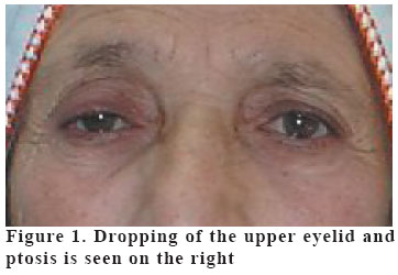
of Nephrology1, Departments of Radiology2 and Pulmonary Diseases3, Van, Turkey Code Number: gm05037 Internal jugular vein cannulation has become the preferred approach for temporary vascular access for hemodialysis. Internal jugular vein cannulation is associated with a high rate of successful catheter placement. However, significant complications such as internal carotid artery (ICA) puncture, vessel erosion, thrombosis and infection can occur. We present one case of Horner's syndrome (without arterial punction) occurring following internal jugular venous cannulation. We suggested that, clinicians need to be aware of the risk of Horner's syndrome as a possible complication of percutaneous hemodialysis catheterization via the internal jugular vein and should avoid repeated manipulations Key words: Horner Syndrome,internal jugular vein canulation,hemodialysis INTRODUCTION Internal jugular vein (IJV) catheterization is commonly used in order to obtain temporary acces to circulation enabling hemodialysis in patients with renal failure; also internal jugular vein catheterization is associated with a high rate of successful catheter placement. However, significant complications such as internal carotid artery (ICA) puncture, pneumothorax, vessel erosion, thrombosis, airway obstruction and infection can occur. The most common complication is ICA puncture. Hemodialysis patients may have to undergo multiple catheter placements and vascular access interventions. This, along with their comorbid conditions, increases the risk of such complications (1-3). Here we report a patient on hemodialysis who developed Horner's syndrome(HS) following right internal jugular vein catheterization . CASE A 70 year-old woman with end stage renal disease was admitted to hospital with metabolic acidosis and uremia. A double-lumen hemodialysis catheter was inserted percutaneously by anatomic landmark technique via right internal jugular vein for emergent hemodialysis after three attempts of cannulation of the vein for guide wire insertion. Because of difficulty in suitable positioning of the patient for cannulation guide wire insertion was possible at the thirth intervention. Dropping of the right upper eyelid and right sided ptosis developed 24 hours after the catheter insertion (see photograph). Ophtalmic examination confirmed HS in right eye with a 3,5 mm ptosis. The size of the right pupil was 2,5 mm and the left pupil was 4,5 mm in the examination room. Both pupils reacted normally to light. Pronounced lack of anhidrosis on the forehead above the right eye was also detected. No other neurologic finding or evidence of mass lesions in the neck or pulmonary apex was present. Radiologic examination including chest radiography, cervical spine radiography, neck CT and chest MRI was normal. The ptosis and anisocoria had partially resolved without any treatment after one month. DISCUSSION Horner's syndrome is characterized by an interruption of the oculosympathetic nerve pathway somewhere between its origin in the hypothalamus and the eye. The classic clinical findings associated with HS are ptosis, pupillary miosis and facial anhidrosis. Other findings may include apparent enophthalmos, increased amplitude of accommodation, heterochromia of the irides (if it occurs before age two), paradoxical contralateral eyelid retraction, transient decrease in intraocular pressure and changes in tear viscosity (1-4). The cause of HS are classified as a pre or post-ganglionic because of the long course of sympathetic innervation to the eye. Common causes of post-ganglionic HS include trauma, cluster migraine headache and neck or thyroid surgery. The common etiologies of acquired preganglionic Horner's syndrome include trauma, aortic dissection, carotid dissection, tuberculosis and Pancoast tumor and performed epidural anesthetic intervention (4-8). In addition, preganglionic HS may be developed shortly after repeated attempts of cannulation of the internal jugular vein (1). In this situation, excessive rotation of the head and neck may have disturbed the normal relationship of the internal jugular vein to the sympathetic trunk and repeated invasive manipulations of the internal jugular vein may have resulted in interruption of the oculosympathetic outflow causing a preganglionic HS (1,9,10). The damage done by catheterization could also be influenced by calibers of catheter. Double-lumen catheters is thicker than the single-lumen catheters, usually needing an additional dilator. In our case, we thought that the causative factors of HS were repeated puncture attempts, thick dilator use, catheter insertion, and microhemorrhage which could not be seen on radiologic examination. For hemodialysis catheter insertion ultrasonographic guidance is recommended in K-DOQI guideline (11). And this method may prevent catheter insertion complications such as HS. In conclusion, in clinical practice, clinicians need to be aware of the risk of HS as a possible complication of percutaneous hemodialysis catheterization via the internal jugular vein and should avoid repeated manipulations. (Figure 1) REFERENCES
Copyright 2005 - Medical Investigations Society |
