 |
| About Bioline | All Journals | Testimonials | Membership | News |
|
||||||
|
||||||
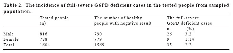
European Journal of General Medicine, Vol. 3, No. 1, 2006, pp. 5-10 INCIDENCE OF SEVERE GLUCOSE-6-PHOSPHATE DEHYDROGENASE (G6PD) DEFICIENCY IN COUNTRYSIDE VILLAGES OF THE CENTRAL CITY OF MANISA, TURKEY Ersin Minareci, Selim Uzunoğlu, Orkide Minareci Celal Bayar University, Faculty of Sciences and Arts, Biology Department, Campus of Muradiye, Manisa, Turkey Correspondence: Selim Uzunoğlu Biology Department, Faculty of Sciences and Arts, Celal Bayar University, Campus of Muradiye, 45030, Manisa/Turkey Phone: 902362412151-118, Fax:902362412158 E-mail: selim@bayar.edu.tr Code Number: gm06002 Aim: The primary objective of this study was to determine the incidence of severe G6PD deficiency in selected countryside villages of central city of Manisa in Turkey. Secondarily to inform and protect G6PD deficient people from acute hemolytic crisis and neonatal jaundice by delivery of the updated protective food and drug list prepared in the light of the WHO- G6PD Working Committe reports. Key words: Severe G6PD deficiency, fluorescence spot test, acute hemolytic anemia, Manisa, Turkey. INTRODUCTION Glucose 6-phosphate dehydrogenase (G6PD, EC 1.1.1.49; locus linked to the X chromosome at the q28 locus) expressed in all tissues, is the first enzyme of pentose phosphate pathway, where 5-carbon sugar, ribose, and NADPH were synthesized by coupled oxidation/reduction reactions (1). The function of the normal G6PD enzyme is critical to human survival since G6PD is the only enzyme producing NADPH required for antioxidative repair systems in circulated erythrocytes (2). G6PD deficiency describes all structural and functional disorders reducing the catalytic function of G6PD protein expressed from G6PD gene. It is the most common human enzyme deficiency in the world. It affects an estimated 400 million people and displays the characteristics of X-linked inheritance (3,4). Severe G6PD deficiency causes several mild to severe health problems either directly or indirectly depending on the conditions. G6PD deficient individuals can expect several clinical manifestations including hemolytic anemia, neonatal jaundice, abdominal and/or back pain, dizziness, headache and dyspnea. If the update and right combination of scientific information and technology was built up and applied, the occurence of health problems would be prevented. The fifteen percentage of normal enzyme activity and its lower values were categorized as severe G6PD deficiency based on the classification of World Health Organization (WHO). This severe form of G6PD deficiency should be screened in populations where the incidence is one percentage and higher. Protective advice should be given to given to severity deficient people (5). The 7.5% of world population are carriers of G6PD deficiency, and 2.9% were G6PD deficient according to WHO reports (6). G6PD deficiency screening studies carried out in Aegean region indicates that the frequency of severe G6PD deficiency in population conforms to the screening criteria of WHO G6PD Deficiency Working Committee. If severe G6PD deficient people consume certain oxidative foods such as fava beans and drugs, an acute hemolytic anemia can be induced in affected individuals especially in children and adults. If the cause of acute hemolytic anemia developing with jaundice could not be find in first 12 hours and blood could not be supplied, the person would be exposed to death risk between 18 and 72 hours. It may also lead to neonatal jaundice and related health problems in many G6PD deficient newborns (7, 8). Fortunately when severe G6PD deficient people were diagnosed earlier by screening tests and given updated forbidden food and drug list, the health problems related to the G6PD deficiency could be definitely prevented. Having considering that there is no sign of the disease in normal conditions but leading to the deathly events conditionally, it makes the G6PD deficiency screening studies very important and beneficial in population level. The primary objective of this study was to determine the incidence of severe G6PD deficiency in selected countryside villages of central city of Manisa in Turkey. Secondarily to inform and protect G6PD deficient people from acute hemolytic crisis and neonatal jaundice by delivery of the updated protective food and drug list prepared in the light of the WHO- G6PD Working Committe reports. MATERIAL AND METHODS The blood samples were collected from non-related 1604 people (male 816; female 788) living in some countryside villages of central city of Manisa. The size of living population in the country side villages of central city of Manisa is about 8672 (9). The severe G6PD deficiency screening study were carried out in the villages given in Table 1. The geographic locations of the villages where the tested people live were given in Figure 1. The males and females were randomly screened in the villages. There was no gender bias in this study. The blood samples were collected only from healthy people. The diagnosis of severe G6PD deficient cases was made preferentially by using standardized home made fluorescence spot test developed by Uzunoglu S instead of commercial Beutler’s fluorescence spot test (10). The preferred and used screening test were modified and standardized according to guidelines of International Hematology Standardization Committee and Sigma G6PD standards with normal (non-deficient) (9-12 U gr/Hb) and patient (full-deficient) controls (0.0- 0.4 U gr/Hb). Blood samples from finger tips for each person were collected into heparinized eppendorf tubes and were stored in ice-boxes. They were kept in +4 oC until fluorescence spot test were run. The modified protocol of Beutler’s Fluorescece spot test was performed (10). Five ml blood from each sample were delivered into each eppendorf tube containing 45 ml spot test reagent. 10 ml samples taken out from spot test reactions were dropped separately in whatman paper with one size after 10 minutes of incubation at room temperature. Dried spots containing the mixture of hemolysate and spot test reagent were evaluated directly by looking at the spot colour under the ultraviolet light (366 nm wavelength) in dark room. In this test, the blood samples with both normal G6PD activity and mild deficiency appear in heavy green fluorescence colour based on the amount of hemoglobin in the blood sample. This greenish colour was interpreted as negative result in terms of severe G6PD deficiency and normal healthy person. If dried completed test reaction spots were in heavy brownish colour, this was accepted as positive result in terms of severe-full G6PD deficiency. Three dilutions of positive full-deficient blood samples were reassayed in triplicate in order to avoid the possible false-positive results due to high concentrations of hemoglobin. RESULTS Thirty-five out of 1604 people screened by spot test were found to be severe G6PD deficient. The prevalence of severe G6PD deficiency was 2.2% in sampled population(Table 2). The distribution of severe deficient G6PD cases in males and females was as below: twenty-six out of thirty-five cases was male. Only nine out of thirty-five cases was female. The percentage of severe deficient patients was 74.3% in males and 25.7% in females. There was also marked differences in prevalence between males (3.22%) and females (1.14%). The difference was statistically significant (p<0.001). The distribution of thirty-five severe G6PD deficient cases in villages were displayed in Table 3. The high numbers in positive cases were found in Ucpinar (10), Yenikoy (9) and Horozkoy (7) villages. There were no positive cases in the villages of Akgedik and Karakılınçlı with mountainous geography and high altitude (Table 3). DISCUSSION Fluorescence spot test has the highest validity and specificity in the diagnosis of severe G6PD deficiency for both homozygote males and females. Moreover it was standardized by International Hematology Standardization Committee, and its use in screening severe G6PD deficient cases was advised by WHO-G6PD group (6,10). Therefore the Florescence spot test was preferred in qualitative screening of severe-full G6PD deficient cases in order to determine the incidence of severe G6PD deficiency in the villages of central city of Manisa. It was not necessary to determine quantitatively the activity of G6PD enzyme since the aim of study was to find the severe G6PD deficient cases in the sampled population. Before using the spot test, the mixture of test reagents, prepared manually, were calibrated by using Sigma G6PD control- normal (Sigma G–6888) and G6PD control–patient (severe-full deficient) (Sigma G–5888) standards. In the evaluation of fluorescence spot test findings, the colour of spot test made by Sigma standards were used as a reference. The heavy green colour indicated the G6PD non-deficient blood sample, while the light and heavy brown colour was the indicator of full-severe deficient blood samples. In order to prevent the false positive results among the positively diagnosed blood samples, dilution gradient of blood samples were studied and the differences in colour formation were optimized (11,12). Thirty-five out of 1604 people screened by spot test were found to be severe G6PD deficient. The prevalence of severe G6PD deficiency was 2.2% in sampled population. The incidence was above the 1% in population based on WHO-G6PD group criteria of inherited disease significance level.The percentage of full-severe deficient patients was 74.3% in males and 25.7% in females.There was also marked differences in prevalence between males (3.22%) and females (1.14%) in sampled population. This remarkable difference conforms the X linked inheritance of G6PD deficiency. The high numbers in positive cases were found in Ucpinar (10), Yenikoy (9) and Horozkoy (7) villages. There was remarkable difference in the number of positive cases compared the other villages. There were no positive cases in the villages of Akgedik and Karakılınçlı with mountainous geography and higher altitude. The locations of villages with more cases were close to Gediz river and delta. The high incidence of severe G6PD deficiency in the sampled Turkish population may be due to the geographic region with the history of malaria endemic and was surrounded by Gediz river and several waterlands. There were few studies on the incidence of severe G6PD deficiency in Aegean region. In one study made in Manisa by Demircan K (1999) in newborns and adults (n=601), the activity of G6PD enzyme were measured quantitatively and the incidence of severe G6PD deficiency were found to be 2.82 %. The incidence in males and females were 5.37 % and 0.62 % respectively (13). The incidence of our sampled population (2.2 %) was quite close to the data(2.82 %) obtained by Demircan K. In one qualitative screening study (n:1950) by using fluorescence spot test, the prevalence of severe G6PD deficiency were found to be 1.23% in the province of Denizli (14).
In another study (1985) by using 500 chord blood samples, the incidence of severe G6PD deficiency was revealed to be 2.4% in Antalya province located in the mediterranean cost of Turkey (15). Our severe G6PD prevalence data (2.2%) was similiar to the data in the mentioned literature and confirms that severe G6PD deficiency is quite high in Aegean region. Moreover, the regional people consume the fava beans in seasonal diet composition. There are many drugs that may induce oxidative stress leading to hemolytic crisis in severe G6PD deficient people. Therefore there is a need for screening for G6PD in the region and inform them about the disease and the list of food and drugs that may lead to acute hemolytic crisis. In conclusion, this high incidence of severe G6PD deficiency implies that this inherited metabolite disorder is an important health problem in Manisa region and it is necessary to carry out large-scale screening in the whole population since severe G6PD deficiency related health problems are preventable. For this reason it must be included in the spectrum of genetic screening tests in regional health policy.
ACKNOWLEDGEMENT: This study is the summary of master thesis of Ersin Minareci under the supervisor of Selim Uzunoğlu submitted to The Institute of Natural Sciences. Celal Bayar University, Manisa. 2000. REFERENCES
Copyright 2006 - Medical Investigations Society |
