 |
| About Bioline | All Journals | Testimonials | Membership | News |
|
||||||
|
||||||
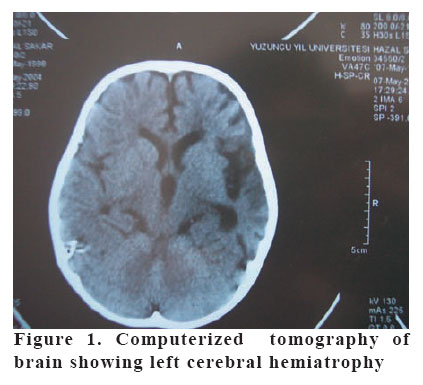
European Journal of General Medicine, Vol. 3, No. 1, 2006, pp. 32-34 ALSTRÖM SYNDROME ASSOCIATED WITH CEREBRAL INVOLVEMENT: AN UNUSUAL PRESENTATION Cahide Yılmaz1, Hüseyin Çaksen1, Nebi Yılmaz2, Ahmet Sami Güven1, Derya Arslan1,Yaşar Cesur3 Yüzüncü Yıl University, Faculty of Medicine, Departments of Pediatric Neurology1, Neurosurgery2 and Pediatric Endocrinology3, Van, Turkey Correspondence: Cahide Yılmaz, MD Yüzüncü Yıl University, Faculty of Medicine, Department of Pediatric Neurology, Van/Türkiye Tel: 904322176128 Fax: 904322150479 E mail: cahideyilmaz@yyu.edu.tr Code Number: gm06007 Alström syndrome (AS) is a rare autosomal recessive disorder, characterized by retinal degeneration, progressive hearing impairment, truncal obesity and non-insulin dependent diabetes mellitus. A 6-year-old girl was admitted with aphasia, deafness, strabismus, abdominal distention, and weakness on the right body side. The physical and laboratory examination revealed psychomotor retardation, right hemiparesis, sensorioneural hearing loss, aphasia, eye and teeth abnormalities, hyperpigmentation, truncal obesity, hepatosplenomegaly, severe iron deficiency anemia, delayed bone age, and cerebral hemiatrophy. Based on these abnormal findings she was diagnosed as AS. According to our knowledge this is the first case of AS with cerebral involvement. This last finding may be a component of the syndrome. Key words: Alström syndrome; brain involvement, case report INTRODUCTION Alström syndrome (AS) is a rare autosomal recessive inherited disorder (1), characterized by retinal degeneration, progressive hearing impairment, truncal obesity and non-insulin dependent diabetes mellitus. It may also include acanthosis nigricans, alopesia, short stature, cardiomyopathy, abnormal liver and renal functions, bone-muscle disorders and metabolic and endocrinological abnormalities (1, 2, 3, 4). Since the first description of the syndrome in 1959, there have been fewer than 100 reported cases in the world (3). In this paper, we reported a case of AS with left cerebral hemiatrophy. To our knowledge this is the first case of AS with cerebral involvement in the literature. CASE A 6-year-old girl was admitted with aphasia, deafness, strabismus, abdominal distention, and weakness on the right body side. She had aphasia and deafness since the birth. Strabismus/abdominal distention and right hemiparesis were first noted three years and three months before admission to the hospital, respectively. The patient was born by vaginal delivery after a 40-week uncomplicated pregnancy, but her developmental milestones were retarded. She began to hold the head on the 10th month of age and sit without support on the 18th month of age and to walk on the 3rd year of age. There was a second degree of consanguinity between the parents. She had healthy three brothers and three sisters. However, her one younger sister who had similar abnormal findings including congenital aphasia and deafness, generalized hyperpigmentation on skin and truncal obesity died when she was 3-year-old. The weight and height were 19 kg (25-50 percentile) and 101 cm (below the 3rd percentile), respectively. The body mass index was 18.8 kg/m2. The physical examination showed generalized hyperpigmentation on skin, truncal obesity, mild psychomotor retardation, right hemiparesis, sensorioneural hearing loss and aphasia, but no acanthosis nigricans or polydactyly. An ophthalmologic examination documented hypermetropia, alternate esotropia and bilateral peripheral retinal degeneration. Gingivostomatitis, flattened papilla on the tongue, and yellow-brown discolored enamel bands of the anterior teeth were noted. Cardiac examination showed II/VI degree systolic murmur on the mesocardiac focus. She had five centimeters hepatomegaly and two centimeters splenomegaly. Deep tendon reflexes were brisk on all extremities. The remaining of the physical findings were normal. Laboratory investigation revealed normal urinary analysis. Hemoglobin was 2.9 g/dL; leukocyte count 5,740/mm³; platelet count 380,000/mm³; reticulocyte count <%1; and erythrocyte sedimentation rate was 27 mm/h. Serum electrolytes, renal and liver function tests including serum triglyceride, cholesterol and ceruloplasmin were normal. Serum iron level was 15 µg/dl (N: 22-184 µg/dl); iron binding capacity >699 µg/dl (N: 250-400 µg/dl); and ferritine <1.5 ng/ml (N: 7-140 ng/ml). Serum thyroid function tests, adrenocorticotrophic hormone and prolactine levels, and glucose tolerance test were all within normal ranges. Serum insulin level was 56.6 µIU/ml (N: 6-27 µU/ml) and C-peptide >7 ng/ml (N: 0.9-4 ng/ml). Serum insulin growth factor-binding protein-3 was 1.2 µg/ml (N: 1.3-5.6 µg/ml) and insulin growth factor-1 level was 12.4 ng/ml (N: 52-297 ng/ml). Growth hormone response to clonidine and L-Dopa stimulating tests was found to be decreased. Hepatitis markers, viral serology including TORCH, Ebstein-Barr virus and parvovirus and salmonella and brusella agglutination tests were unremarkable. Serum immunoglobulin levels were normal. Bone marrow examination was consistent with iron deficiency anemia. The result of blood and urine cultures was negative. Her bone age was four-year-old. Thorax radiography and echocardiographic examination was normal. Abdominal ultrasound showed hepatosplenomegaly. Computerized tomography (CT) of brain showed left cerebral hemiatrophy (Figure 1). Brain stem auditory response could not be performed, unfortunately. Based on the above mentioned clinical and laboratory findings, the patient was diagnosed as AS. She was discharged from the hospital without treatment, but genetic counseling was given to the parents. DISCUSSION The molecular basis of the insulin resistance in this disease is not understood. Investigators have reported normal insulin-mediated RNA synthesis in the patients, suggesting a postreceptor defect in AS (2). Growth hormone deficiency has been reported in AS (5). It has been suggested that resistance to several polypeptite hormones including insuline, vasopressin, gonadotropins and growth hormone might be responsible for some aspects of the disease (2). The basic pathology of retinal degeneration in this syndrome is open to debate (2). All reported patients usually have nistagmus by one year of age and photophobia is common. Vision usually is severely impaired in patients younger than one year of age, and the symptoms may be confused with those of Leber’s congenital amaurosis or achromatopsia. The patients with AS cited in the literature generally have a visual acuity of less than 6/60 by 10 years of age(5). Liver involvement in Alström syndrome was first described by Connolly et al. Their 11-year-old patient developed hepatic dysfunction at age 8 (6). No viral, autoimmune, or metabolic etiology was idendified (7). Multiorgan involvement, as seen in AS, is characteristic of mitochondrial disorders. In previous reports, AS showed no ultrastructural mitochondrial abnormalities, but slight mitochondrial pleomorphism in a liver biopsy. This does not exclude all primary mitochondrial disorders, especially those related to enzyme defects in the pathways different from the respiratory chain (8). Our patient had normal liver functions, but hepatosplenomegaly was present. Generally, moderate sensorineural deafness is detected within the first decade (2). In the literature, one study reported onset of hearing defect at 5 to 12 year of age (5). Individuals with this syndrome may resemble some other genetic disorders, like Laurence-Moon or Bardet-Biedl syndrome, with some common characteristics such as obesity, hearing loss, retinitis pigmentosa, and age of blindness onset (2). Our patient had deafness; however brain stem auditory response could not be performed. Mental functions were stated to be normal in previously reported cases of Alström syndrome (2). Evaluating intelligence in individuals with deafness is difficult, and pseudo-retardation might be detected due to other handicaps. In the literature oral findings were described in two cases of AS. In both cases, gingivitis was present and also light yellow-brown discolored enamel bands were observed on the anterior teeth (9). Our patient had flat tongue papilla, bad mouth hygiene, gingivitis and yellow-brown discolored enamel bands on the anterior teeth. Previous studies have reported no information regarding brain findings. Our patient had weakness on the right hand, and cranial CT revealed hemiatrophy on left hemisphere of the brain which was not previously described. Multiorgan involvement, as seen in AS, is characteristic of mitochondrial disorders; however in previous reports, no ultrastructural mitochondrial abnormalities were shown in liver and heart biopsies from patients with AS (8). A multidisciplinary approach to clinical management is clearly required from the time of diagnosis of AS. This is particularly crucial in detection, evaluation and treatment of several important components of the syndrome. In conclusion, there is no information about brain involvement in Alstrom syndrome in previous reports. This is the first reported case of Alström syndrome with cerebral hemiatrophy with an unusual involvement which may also be a coincidental association. REFERENCES
Copyright 2006 - Medical Investigations Society The following images related to this document are available:Photo images[gm06007f1.jpg] |
| |||||||||
