 |
| About Bioline | All Journals | Testimonials | Membership | News |
|
||||||
|
||||||
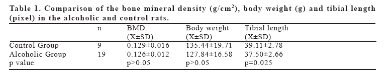
European Journal of General Medicine, Vol. 3, No. 2, 2006, pp. 54-57 THE EFFECTS OF ALCOHOL EXPOSURE DURING INTRAUTERINE AND POSTNATAL PERIOD ON BONE MINERAL DENSITY AND BONE GROWTH AND BODY WEIGHT IN RATS’ VIRGIN OFFSPRING Kadir Ertem1, Ersoy Kekilli2, Nurzat Elmali1 , Feti Ceylan1 Inonu University, Faculty of Medicine, Departments of Orthopedics and Traumatology1 and Nuclear Medicine2 , Malatya, Turkey Correspondence:Kadir Ertem, MD. Inonu University, T. Ozal Medical Center, Department of Orthopedics and Traumatology, 44315 Malatya, Turkey. Tel: 904223410660/5110, Fax: 904223410728 E-mail: kertem@inonu.edu.tr Code Number: gm06012 Aim: To investigate the effects of alcohol contained continuous modified liquid diet ingestion in rats’ offspring on bone length, bone mineral density and body weights. Key words: Alcoholism, Intrauterine growth retardation, Bone mineral density. INTRODUCTION It has been recognized that maternal heavy alcohol consumption during pregnancy may detrimental effects result to the developing fetus. Jones et al (1975) summarized these developmental defects as fetal alcohol syndrome (FAS) that its characteristics including reduced body growth, brain and behavioral dysfunction, and anomalies in craniofacial structure(1). The studies in human and animal models revealed that growth retardation was shown as decreased body length or short stature. These growth deficits do not recover in postnatal period and have been demonstrated to persist till 14 years of age in humans (2, 3). Excessive consumption of alcoholic beverages is associated with increased bone resorption, decreased bone formation and mild mineralization defect (4-8). The mechanisms through which alcohol induces bone loss are currently not well understood. Some authors were emphasized as alcohol ingestion induces osteoclastogenetic changes and bone loss (9,10). Quantitative computed tomography and quantitative magnetic resonance imaging provide an accurate measurement of the three-dimensional geometry of bone and its trabecular bone compartment (11, 12). Because of their costs, required time and radiation dose, dual-energy x-ray absorptiometry (DEXA) is currently the most widely used tool for the measurement of bone mineral content because of its accuracy, precision, stability, and low dose of radiation as well as the speed and ease of scanning (11,13). In this study, we aimed to analyze bone mineral density (BMD) changes at the total tibia in addition to longitudinal bone growth and body weight in alcoholic and non-alcoholic rats’ offspring. MATERIAL AND METHODS Animals The 19 of 27 offspring of 4 Alcoholic female rat as Alcoholic group and the 9 of 15 offspring of non-alcoholic female rat as Control Group were admitted to this study. These rats were fed by modified liquid diet without ethanol till 12 weeks of age after weaning at 4 weeks age. All rats and their young offspring were placed in same living conditions (12 hours of light and 12 hours of darkness; mean room temperature 20-22°C and 50% environmental humidity) and habitat. Animals had free access to both diet and drinking solution throughout the experimental period of pregnancy and lactation period. All experiments in this study were performed in accordance with the guidelines for animal research from the National Institutes of Health (National Research Council, 1985) and were approved by the Committee on Animal Research at Inonu University Medical Faculty, Malatya, Turkey. Diet Alcohol-provided female rats (n=4) were individually housed and were submitted forced alcoholization as previously described method (14). Briefly, all rats were fed by a modified liquid diet (MLD) for 7 days. The composition of the MLD was cow’s milk, 925 ml; vitamin A, 5000 IU; and sucrose, 17 g. The MLD had 4.187 kj/liter. At the end of 7 days, MLD with 2.4% ethanol (v/v; ethanol 95.6%, Tekel, Turkish State Monopoly, Izmir) was administered to Alcoholic group (n=19) for 3 days. Then, the ethanol concentration was increased to 4.8% for 3 days, and finally the rats completed the rest of the 8 weeks with a diet containing 7.2% ethanol. When the ethanol concentration was increased, sucrose was reduced to maintain the isocaloricity of the diet. These rats were conceived at the end of this period. Pregnant females were replaced individually in their cages and assigned again to 7.2% ethanol in tap water as the sole source of liquid with food “ad libitum”, during the pregnancy and lactation period. Pair-fed control female rats (n=2) were fed an isocaloric MLD without ethanol throughout the experiment. The liquid diet was offered in special glass bottles to prevent spillage. Water was also freely accessed in different bottles. All rats were maintained on the liquid diet regimen for 12 weeks. At the end of the experiment, the weights of the rats were recorded. These rats were conceived at the end of this period. Pregnant females were replaced individually in their cages and assigned again to isocaloric MLD without ethanol during the pregnancy and lactation period. Surgical Technique At the postpartum 90th days, all rats were sacrificed and the tibias were preserved without skin in 10% formaldehyde solution. Bone Densitometric Investigation In the all rats, bone mineral density (BMD) of the bilateral tibia were measured with dual-energy X-ray absorptiometry (DEXA) (QDR 4500/W, Hologic Inc., Bedford, MA, USA) and results were evaluated by the same examiner. DEXA equipment uses switched pulsed stable dual-energy radiation with 70 kV and 140 kV. Subregion analysis software was used to analyze of the whole tibia (15). BMD value of the tibia was used, in g/cm2. All tibia length was obtained as pixel values by bone mineral densitometry. Data Analysis Statistical analyses were performed with version 11.0 of SPSS for Windows (SPSS Inc., Chicago, IL). BMD, body weight, and tibia length of each group were expressed as mean ± SD and were compared by use of the Mann-Whitney U test for detecting differences between independent groups. A value of p< 0.05 was considered to be statistically significant. RESULTS There was an approximately 2.5% lower BMD in chronic alcohol provided offspring (0.126±0.012) than the non alcohol provided ones (0.129±0.016), but not significant (p>0.05) (Table I, Figure 1). There was not any significant difference according the body weights between alcoholic mother and non-alcoholic offspring (p>0.05). However there was a significant fall in body length in offspring which maternal alcohol consumption during prenatal and postnatal period (p<0.05) (Table I). DISCUSSION Metabolic bone disease, mainly osteoporosis, is frequent in alcoholics. Despite its pathogenesis seems to be multifactorial. Santolaria et al found that alcoholic osteopenia may be interpreted as a form of nutritional osteoporosis in their cross-sectional study in 181 male alcoholic patients. The most significant bone loss was evident in patients who have very irregular feeding habits (16). However, Norton et al emphasized the detrimental effects of alcohol on growth retardation, facial dysmorphology, immune system deficiencies, endocrine dysfunction, and central nervous system alterations during prenatal period that obtained from experimental studies (17). Despite the body weight and BMD measurements were not affected significantly in Alcoholic group, but the longitudinal growth. There is not several studies related the effect of maternal alcohol consumption on maternal and fetal calcium homeostasis and fetal skeletal development. Keiver et al found hypocalcemia in pregnant rats with feeding by alcoholic diet at five weeks but placental calcium transfer to fetus was not impaired. On the day 21 of gestation, the collected fetuses of maternal ethanol consumption group were found decreased fetal growth and delayed fetal skeletal development (18). However with their findings that fetal skeletal ossification was not severely affected when maternal consumption occurred for only three weeks and concluded as skeletal growth impairment was improving severely with increasing duration of maternal alcohol consumption (19). Murillo-Fentes et al found that chronic alcohol administration during gestation and/ or lactation detrimentally effects pup growth at weaning as indicated by its effect on milk consumption, pup and organ weights (20). In present study, there was not any difference between non-alcoholic offspring and alcoholic mother offspring according the body weights (p>0.05). This may be related the consumption of isocaloric diet regime in both group. However there was a significant diminution in body length in offspring which maternal alcohol consumption during prenatal and postnatal period (p<0.05). Healthy bones in the adult and elderly people are dependent especially on the development, during the intrauterine period and postnatal younger years of a healthy bone structure and adequate peak bone mass. Day et al demonstrated in their cohort study that the growth deficit due to prenatal alcohol exposure was continuing at age 14 years (2). In this study, despite we found 2.5% diminution of BMD in offspring who had treated alcohol during intrauterine and postnatal period but it was not found statistical significant (p>0.05). The handicaps of this study are the lack of biochemical markers analyses for natal and postnatal calcium homeostasis and analyses of histomorphometric changes in bone stature in these rats. In conclusion, our findings of mild but non-significant loss in tibia BMD and body weights in offspring rats of treated alcohol during intrauterine life may be attributed the irregular feeding habits due to social factors in chronic alcohol consumption in human. Our findings related the significant short tibia length in chronic alcohol treated during prenatal and postnatal period are found correlated with the studies in human and animal models revealed that growth retardation was evident with decreased body length or short stature. REFERENCES
Copyright 2006 - Medical Investigations Society The following images related to this document are available:Photo images[gm06012f1.jpg] [gm06012t1.jpg] |
| |||||||||
