 |
| About Bioline | All Journals | Testimonials | Membership | News |
|
||||||
|
||||||
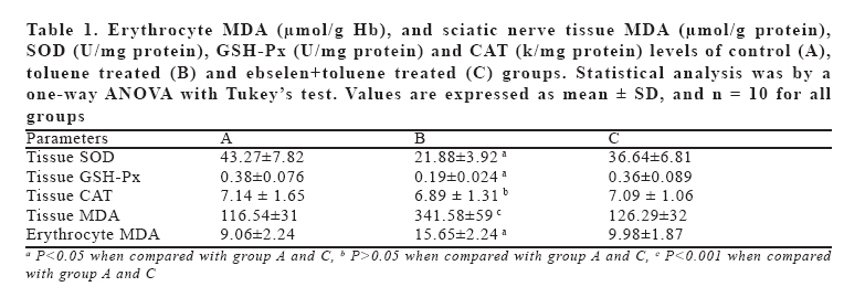
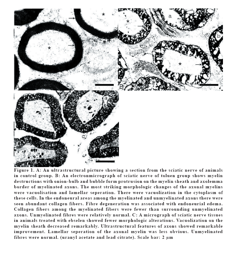
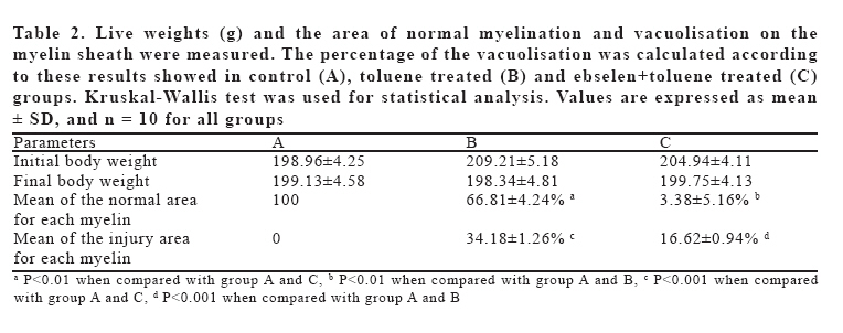
European Journal of General Medicine, Vol. 3, No. 2, 2006, pp. 64-72 EBSELEN PROTECTS AGAINST OXIDATIVE AND MORPHOLOGICAL EFFECTS OF HIGH CONCENTRATION CHRONIC TOLUENE EXPOSURE ON RAT SCIATIC NERVES Ömer Coşkun1, Mehmet Yüncü2, Mehmet Kanter1, Sadık Büyükbaş3 Trakya University, Faculty of Medicine, Department of Histology and Embryology1, Edirne, Gaziantep University, Faculty of Medicine, Department of Histology and Embryology2 Gaziantep, Selcuk University, Faculty of Medicine, Department of Clinical Biochemistry3, Konya, Turkey Correspondence: Dr. Mehmet Kanter, Trakya Üniversitesi, Tıp Fakültesi, Histoloji-Embriyoloji AD, Edirne-Türkiye Fax: 902842352730, Tel: 902842357641 E-mail: mkanter65@yahoo.com Code Number: gm06014 Aim: Neuroprotective effects of ebselen against free radical damage have been studied extensively. Toluene generates reactive oxygen species (ROS) and the toxic effects relating to these reactants. The aim of this study was designed to investigate the effects of chronic toluene inhalation in high concentration on lipid peroxidation, antioxidant enzyme activities and ultrastructural changes in the sciatic nerves of rats. Key words: Toluene, ebselen, lipid peroxidation, antioxidant enzymes, ultrastructural, sciatic nerve INTRODUCTION Toluene is a organic solvent heavily used in the rubber, plastics, leather, paint, printing and chemical industries (1). It is readily absorbed from the gastrointestinal and respiratory tracts, and to a lesser degree through the skin. Toluene is distributed throughout the body, with accumulation in tissues with high lipid content (2). In humans and animals, the primary effect associated with inhalation exposure to toluene is central nervous system (CNS) depression (3,4). Several studies, carried out with animals, reported an uptake of inhaled toluene in blood and brain (2,5). Toluene’s affinity for the lipid-rich structures of nervous tissue results in CNS toxic effects within minutes (2,6). Despite the widespread abuse of toluene and other solvents, the molecular sites of action of these compounds are largely unknown (7). More is known about the behavioral effects of these substances: toluene and other abused solvents produce effects similar to that of central nervous system depressants. These effects include psychomotor impairment (8), excitation and then inhibition of locomotor activity (9), and loss of righting reflex and sedation (10). In addition, peripheral nerve dysfunction has been reported (11). Mattia et al. (12) demonstrated that intraperitoneal injection of toluene caused a significant elevation in the rate of reactive oxygen species (ROS) generation and a reduction in glutathione (GSH) levels in the brain. Long-term toluene abuse has led to neuropsychiatric and neurobehavioral disorders, which in many cases, but not all, were reversible. Some chronic toluene abusers have developed structural CNS damage (3). Volatile substances containing toluene are frequently abused by young people, thus leading to neurological damage. In fact, glue sniffing is frequent among secondary school children and young adults because it is more easily available and cheaper than other drugs (13). Ebselen, 2-phenyl-1,2-benzisoselenazol-3[2H]-one, is a lipid-soluble seleno-organic compound that potently inhibits lipid peroxidation through a glutathione peroxidase-like activity (14). Also, recent animal studies show neuroprotective, antioxidant, and anti-inflammatory actions of ebselen (15,16). But to date, no study reported the effect of ebselen on biochemical and histopathological changes due to toluene exposure on rat sciatic nerves. In the present study, it was aimed to investigate the possible beneficial effects of ebselen on lipid peroxidation, antioxidant enzyme activities and histopathological changes of chronic toluene exposure on rat sciatic nerves. MATERIAL AND METHODS Animals Thirty male Wistar albino rats, weighing 150-250 g, were alloted in three experimental groups: the control group (A), toluene treated group (B) and ebselen+toluene treated group (C); n=10 for each group. The animals were purchased from a local supplier (Ankara Laboratories), and housed in individual cages (360 x 200 x 190 mm) 1 month before the start of the experiments. Food and tap water were available ad libitum. In the windowless animal quarter automatic temperature (22±2ºC) and lighting controls (light on at 07 AM and off at 09 PM: 14 h light/10 h dark cycle) was performed. Humidity ranged from 50% to 55%. All animals received human care according to the criteria outlined in the “Guide for the Care and Use of Laboratory Animals” prepared by the National Academy of Sciences and published by the National Institutes of Health. Solvent exposures Animals in group B and C were exposed to 3000 ppm toluene (Merck, 99.5%) in inhalation chambers equipped with a trap, and designed to sustain dynamic and adjustable airflow (11250 mg/m3). This animals were housed in individual cages within an inhalation chamber for 8 h/day (from 08 AM to 04 PM). This application was continued 6 day/week for a total of 16 weeks. Toluene was vaporized by bubbling additional airflow through a flask containing the test compound. The solvent concentration in the chambers was continuously monitored using a gas chromatography (GC). Initial of study there were 12 rats in the toluene group. Toluene dose was very high, so 2 animals died from toluene group during experiment. Control group (A) animals were housed in identical chambers ventilated with fresh air. All rats were weighted at the beginning and after the study. Drug preparation and sample collection Group A received 1ml serum physiologic and ebselen was given i.p. (10 mg/kg, Sigma, St. Louis, MO, USA) to Group C just after toluene exposure per day. At the end of the solvent exposure, all animals were anesthetized with i.p. injection of sodium thiopenthal (100 mg/kg, Sigma, St. Louis, MO, USA) for blood and sciatic tissue harvesting. Twenty minutes later, the anesthetized rats were sacrificed. Animals were pinned onto a dissection board and the peritoneal cavity was opened. Thus, immediately blood samples were collected by cardiac puncture using heparinised syringe and sciatic nerves were rapidly removed. Samples of the sciatic nerves were stored at -40 ºC until the analysis for superoxide dismutase (SOD), glutathione peroxidase (GSH-Px), catalase (CAT) and malondialdehyde (MDA). Blood samples were taken into heparinized tube. Leukocytes and plasma components were seperated by centrifugation of the blood. Erythrocytes were washed three times with 0.9% NaCl solution and packed. Erythrocytes were then stored at -70 °C until study. Biochemical procedures The tissues were homogenised in buffers by means of Ultra Turrax T25 homogenisator. The soluble fraction was prepared by centrifugation at 6000g for 10 minutes. The blood and sciatic nerves were assayed for toluene by gas chromatography (GC). Given the time course of uptake and elimination of toluene in blood and nerve established by Benignus et al. (2). Levels of MDA were determined by the double heating method of Draper and Hadley (17). The principle of the method is spectrophotometric measurement of the colour produced during the reaction to thiobarbituric acid (TBA) with MDA. For this purpose, 2.5 ml of 100 g/l trichloroacetic acid solution was added to 0.5 ml erythrocyte or tissue homogenate in each centrifuge tube and placed in a boiling water bath for 15 min. After cooling in tap water, the mixture was centrifuged at 1000 g for 10 min, and 2 ml of the supernatant was added to 1 ml of 6.7 g/l TBA solution in a test tube and placed in a boiling water bath for 15 min. The solution was then cooled in tap water and its absorbance was measured using a Shimadzu UV-1601 (Japan) spectrophotometer at 532 nm. The concentration of MDA was calculated by the absorbance coefficient of MDA-TBA complex 1.56 × 105 cm-1 M-1, and were expressed in µmol/g Hb erythrocyte and µmol/g tissue protein. Tissue SOD and GSH-Px activities were measured with diagnostic kits by using a Shimadzu UV-1601 spectrophotometer. SOD activity was measured according to the protocol of the method of Sun et al. (18) using the Ransod kit (Randox Laboratories GmbH, Deutschland). This method is based on the ability of SOD to prevent the formation of formazane from 2-(4-iodophenyl)-3-(4-nitro)-5-phenyltetrazolium chloride by superoxide radicals generated by xanthine oxidase/xanthine. The formation of formazane was recorded spectrophotometrically at 550-nm wavelength. Inhibition by 50% of the 2-(4-iodophenyl)-3-(4-nitro)-5-phenyltetrazolium chloride reduction after addition of the sample (0.01-0.02 mg tissue/mg test volume) was defined as 1 U of SOD. The activity of tissue glutathione peroxidase (GSH-Px) was determined by spectrometry with the use of Ransel kit (Randox Laboratories GmbH, Deutschland) and method described by Paglia and Valentine (19). Tissue CAT activity was determined by according to Aebi’s method (20). The principle of the assay is based on the determination of the rate constant (s-1, k) or the H2O2 decomposition rate at 240 nm. In the samples tissue, protein measurements were made according to Lowry’s method (21). Histological assesment Biopsies from the sciatic nerves of the rats were pre-fixed in 3% glutaraldehyde in Sorensen’s phosphate buffer and then post-fixed in 1% osmium-tetraoxide in the same buffer. They were dehydrated in alcohol series and embedded in Spurr’s resin. Semithin sections, which are at 0,6 µm thickness, were stained with Toluidine blue. After the examination of the semithin sections; ultrathin sections were cut at 200 Angstrom. These sections were stained with uranyl acetate and lead citrate. The specimens were examined at Carl Zeiss electron microscopy (EM) 900 and pictures were taken. Image analysis The system used is composed of a PC, hardware and software (Image-Pro Plus 5.0-Media Cybernetics, USA) for image acquisition and analysis, Spot Insight QE (Diagnostic Instruments, USA) camera and optical microscope. The method requires preliminary software procedures of spatial calibration (µm scale) and setting of color segmentation for quantitative color analysis. Five axons from each rat, thus fifty axons for each group, were chosen randomly. The area of normal myelination and the area of vacuolisation on the myelin sheath were measured. Then the percentage of the vacuolisation was estimated. The investigator who performed these measurements were unaware of the experiment. Statistical analysis The data were expressed as mean ± standard deviation (SD) and analysed using repeated measures of variance. Tukey test was used to test for differences among means when ANOVA indicated a significant (P<0.05) F ratio. For the image analysis of the vacuolisation on the myelin sheath data, a nonparametric test (Kruskal-Wallis) was used. Differences were considered statistically significant when the p value was <0.05. RESULTS Initial of study there were 12 rats in the toluene group. Toluene dose was very high, so 2 animals died from toluene group during experiment and the rest of rats had loss of righting reflex and psychomotor impairment. Biochemical findings Erythrocyte MDA, and tissue SOD, CAT, GSH-Px and MDA levels of all groups are shown in Table 1. Toluene exposure significantly increased blood (P<0.01) and tissue MDA (P<0.001) levels, and decreased tissue SOD and GSH-Px (P<0.01), but not tissue CAT (P>0.05) levels. Clearly, in animals treated with ebselen the elevated MDA concentrations in the sciatic nerve due to toluene were largely attenuated compared with those of animals subjected to only toluene (P < 0.01). No difference was observed statistically in the MDA content between control rats and ebselen treated animals (P>0.05). Ebselen treatment restored tissue SOD and GSH-Px enzyme activity back to the control levels (P < 0.01) (Table 1). No difference was observed statistically in terms of blood toluene concentration between group B (40.6±4.8) and A (41.2±4.9). There were also no significant changes in the nerve tissues toluene concentration of group B (54.7±5.8) and C (51.8±5.2). Histological findings Histological examination of the sciatic nerves in control group showed normal structure (Figure 1A). Sciatic nerve electronmicrographs of toluen group shows myelin destructions with onion-bulb and bubble form protrusion on the myelin sheath and axolemma border of myelinated axons. The most striking morphologic changes of the axonal myelins were vacuolisation and lamellar seperation. Shrunken and swollen axons were common. Total damage and deformation of the axons was evident in some nerve fibres. Degenerative changes were also observed in Schwann cells. There were vacuolization in the cytoplasm of these cells. In the endoneural areas among the myelinated and unmyelinated axons there were abundant collagen fibers. Fibre degeneration was associated with endoneurial edema. Collagen fibers among the myelinated fibers were less than surrounding unmyelinated axons. Unmyelinated fibres were relatively normal (Figure 1B). Our results are including myelins, axons and endoneurial degeneration. These results are compatible with grade 3 nerve lesions according to Sunderland and Seddon (22,23). There was no degeneration on the perineurium and epineurium. Histologic evaluation of the tissues in animals treated with ebselen showed fewer morphologic alterations. Myelin breakdown decreased significantly after treatment with ebselen. Ultrastructural features of axons showed remarkable improvement. Vacuolization and lamellar seperation of the axonal myelin was less obvious. The fine structure of Schwann cells was normal (Figure 1C). There were no significant change in the ultrastructure of unmyelinated fibers. The area of normal myelination and the area of vacuolisation on the myelin sheath were measured. The percentage of the vacuolisation was calculated according to these results showed in Table 2. The percentage of the vacuolisation was significantly increased in toluene group. The measurement of the area of vacuolization of the myelin sheath of the axons also showed that there was a significant decrease on the area of the vacuolization of the myelinated fibers after the administration of ebselen (Table 2). Live weights of the rats were not changed significantly in both groups (Table 2). Histopathological findings were correlated with biochemical findings. DISCUSSION Most studies reporting adverse effects due to chronic toluene exposures involve either toluene-containing solvent abuse or occupational exposure to toluene. Solvent abusers are generally exposed to higher levels of toluene than are workers (6,7). Treatment of animals with toluene is known to cause central nervous system injury (3,12). In our study, we showed that chronic toluene inhalation in high concentrations has negative effects on lipid peroxidation and antioxidant enzyme activities and results in ultrastructural changes in the sciatic nerves of rats. A number of case reports have described optic neuropathy caused by toluene (24,25). In fact, toluene exposure produced oxidative stress in several brain areas (26). Acute and chronic effects of toluene on neurons and oxidative stress have been well documented (26), but there is no study regarding the effect of ebselen treatment in terms of prevention of toluene toxicity on peripheral nerves. Distribution was related to blood concentrations and brain lipid content. Studies in the rat and mouse have detected effects of toluene inhalation on brain constituents and morphological and biochemical parameters. High levels approaching 2000 ppm have caused ataxia, prostration and tremors in rats exposed for 7 days (27). At the completion of this 26-week study, there were no treatment-related effects. Regional loss of cells in the hippocampus coupled with abnormal electrical activity in this region was observed when rats were exposed to a toluene concentration of 500 ppm for 8–16 hours/day for up to 5 weeks (28); it was concluded that toluene causes irreversible effects in this area of the brain. Recent experimental studies have been investigated toluene and its metabolites respect with their ROS-enhancing potential in isolated systems and in vivo. GSH levels were depressed in liver and brain regions from toluene-treated rats. In addition, GSH may play an important role in protection against the induction of ROS generation in the central nervous system and liver following exposure to toluene (12). GSH-Px is particularly important antioxidative enzyme in the protection of the brain from damaging effects of ROS. Glial cells contain augmented levels of GSH as well as elevated activities of the enzymes involved in GSH metabolism and higher SOD activity than neurons (29,30). In our study toluene significantly increased the blood and tissue MDA levels, and decreased the tissue antioxidant enzyme activities (SOD and GSH-Px levels), except CAT. These findings may indicate evidence for misbalance of the antioxidant status. Neurons dispose of endogenous antioxidants such as glutathione, vitamins C and E. Neurons also use antioxidant enzymes, although to a less extent than other cell types (31). Amongst these enzymes, GSH-Px reduce hydrogen peroxide and organic peroxides at the expense of glutathione, thereby limiting the production of the highly reactive hydroxyl radical (32). Ebselen is a well-characterized seleno-organic compound with GSH-Px like activity and neuroprotective effects (33). In this study, ebselen administration prevented increase of blood and tissue MDA and decrease of SOD and GSH-Px levels induced by toluene inhalation. Solvent abusers are inhaling very high concentrations of toluene, in some reports exceeding 10,000 ppm in brief episodes (34). Even when toluene exposure conditions in mice (4000 ppm for 8 weeks, intermittently) approximated concentrations inhaled during toluene abuse, there was a lack of histopathological evidence of damage not only in the liver and kidney, but in brain as well (35). Iizumi et al. (36) suggested that chronic toluene exposure (3000 ppm, 4 h/day for 3 weeks) might influence catecholaminergic neural systems. Barroso-Moguel and Villeda (37) reported that inhaling 135 ml toluene (1.5 ml/day for 90 days) caused edema, ballooning and destruction due to lysis in the myelin sheath. In a parallel manner the nerve fibers show edema, irregular thickening and atrophy or disintegration. Baydas et al. (38) investigated the effects of melatonin on the glial reactivity induced by the exposure of rats to the fumes of thinner, which due to the presence of toluene (3000 ppm, for 45 days) are known to be neurotoxic. Pathological studies of the effects of toluene inhalation based on biopsies of the sural nerve have shown swelling of the axons and an extremely thin lamella of the myelin sheath (39). In the present study, chronic toluene exposure caused degeneration of myelins, axons and endoneurial structures. These results are compatible with grade 3 nerve lesions according to Sunderland and Seddon (22,23). There is no single classification system that can describe all the variations of nerve injury. Most of the systems attempted to correlate the degree of injury with symptoms, pathology and prognosis. Oxidative stress and oxidative damage are considered to play a role in the early stages of the pathophysiological processes of specific cancers, cardiovascular diseases and several neurodegenerative diseases (40). ROS including hydrogen peroxide and hydroxyl radical can be generated by ionizing radiation and has the potential to induce diseases (41). Oxidative stress can be prevented by the action of enzymatic and chemical antioxidant defenses. The enzymes that provide the first line of defense against superoxide and hydrogen peroxide include SOD, CAT and GSH-Px (42,43). Ebselen, originally developed as an anti-inflammatory agent (44). Ebselen is effective against membrane hydroperoxides, it inhibits both nonenzymatic and enzymatic lipid peroxidation in cells and has anti-inflammatory activity in various animal models (14,16). In fact, ebselen have been used experimentally and pre-clinically with success in a variety of situations where free radicals were involved (15,16,45). Ebselen has neuroprotective efficacy, as assessed from gray matter damage in experimental models of cerebral ischemia (15). But to date, no study reported the effect of ebselen treatment on toluene induced biochemical and histopathological changes due to induced oxidative damage. In this study, histologic evaluation of the tissues in animals treated with ebselen showed fewer morphologic alterations. Myelin breakdown decreased significantly after treatment with ebselen and also ultrastructural features of axons showed remarkable improvement. The measurement of the area of vacuolization of the myelin sheath also showed that there was a significant decrease on the area of the vacuolization of the myelinated fibers after the administration of ebselen. In summary, we conclude that ebselen therapy causes biochemical and morphologic improvement on sciatic nerves which have been subjected to toluene exposure. We postulate that the neuroprotective effects of ebselen are attributed to its direct and indirect antioxidant actions. We believe that further preclinical research into the utility of ebselen may indicate its usefulness as a potential treatment for toluene-induced injury. REFERENCES
Copyright 2006 - Medical Investigations Society The following images related to this document are available:Photo images[gm06014t2.jpg] [gm06014t1.jpg] [gm06014f1.jpg] |
| |||||||||
