 |
| About Bioline | All Journals | Testimonials | Membership | News |
|
||||||
|
||||||
European Journal of General Medicine, Vol. 3, No. 4, 2006, pp. 148-151 THE EFFECT OF ENDOSULFAN ON LACTIC DEHYDROGENASE ENZYME SYSTEM IN LIVER OF MUS MUSCULUS : A HISTOCHEMICAL STUDY Ergül Belge Kurutaş1, Figen Doran2, Harun Çıralık3 Sütçü İmam University, Faculty of Medicine, Departments
of Biochemistry1 and Pathology3, Kahramanmaras, Çukurova
University, Faculty of Medicine, Department of Pathology2,
Adana, Turkey Code Number: gm06032 Abstract Aim: Endosulfan is an important toxic agent which generates free
oxygen radicals in liver. In Çukurova region, some relation between
the incidence of toxic injury to liver and widespread use of endosulfan has
been suggested. Since endosulfan is a frequently used pesticide, it was planned
to investigate whether endosulfan could do liver injury and increase lactic
dehydrogenase in liver. Key words: Endosulfan, mus musculus, lactic dehydrogenase, enzyme histochemistry
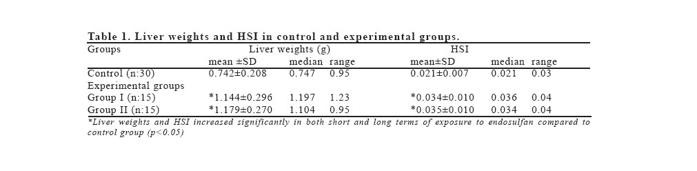
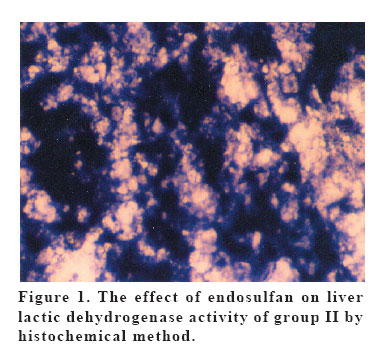
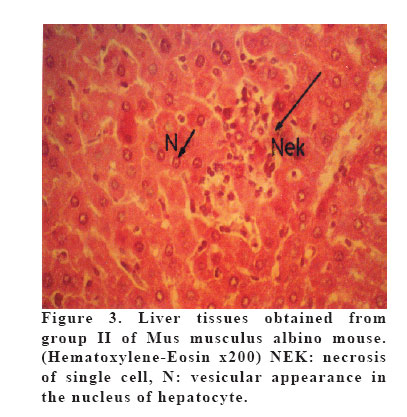
INTRODUCTION Endosulfan is an important environmental pollutant which is a pesticide of organochlorine. Primary site of organochlorine storage in the body is adipose tissue. It is metabolized in the liver as a lipophilic xenobiotic to hepatotoxic intermediates by monooxygenase systems which cause oxidative stress (1). Free radicals generated during oxidative stress cause lipid peroxidation of cell membranes which is in turn prevented by antioxidant enzymes (2-4). Endosulfan alters the activities of lactic dehydrogenase, glucose-6-phosphate dehydrogenase and alkaline phosphatase, and decreases mitochondrial energy production in mice (5). Lactic dehydrogenase (LDH; 1.1.1.27) is a hydrogen transferring enzyme that catalyzes the oxidation of L- lactate to pyruvate with the mediation of NAD+ as hydrogen acceptor. LDH activity is present in all cells of the body and is invariably found only in the cytoplasm of the cells (6). In Cukurova region, some relation between the incidence of toxic injury to the liver and widespread use of endosulfan has been suggested but we have not founded any paper, about the widespread use of endosulfan in Çukurova region. We could not find any study on this subject in the literature. Liver histology and LDH enzyme was investigated in mice (Mus musculus) exposed to endosulfan by enzyme histochemical method. MATERIALS AND METHODS Animals Sixty mature non-inbred M. musculus albino, weighing between 23 to 40 g were obtained from the Medical Sciences Experimental Research Center of the University of Çukurova. They were fed with a standard laboratory diet and tap water. Illumination was 12 hours light/dark cycle and room temperature was 22-24oC. Both experimental and control groups consisted of thirty apparently normal M. Musculus. The experimental group was further divided into two groups (Group I and II) and each group was exposed to endosulfan by oral administration (0.24 mg per 100 g body weight) daily for a 90-day (short term) and 180-day (long term) period. At the end of this period, animals were sacrificed and their livers were quickly removed and cut with a thickness of five micrometers with cryostat sectioning. We followed the Guide for the care and use of laboratory animals. Besides, we obtained approval from ethic commitee Histopathology Liver tissues was fixed in 10% formaldehyde and processed routinely. They were embedded in paraffin. Five µm sections were obtained, stained with Harris hematoxylene-eosin and examined under light microscope (7). Enzyme Histochemistry LDH was evaluated histochemically in the group I and II. Lactic Dehydrogenase Standard Technique (7). Fixation: Unfixed cryostat sections 5-7 micrometers. I. Sections were incubated in appropriate incubating solution at 37°C for 30-60 minutes. II. Then transferred into 15 % formal saline for 15 minutes. III. Washed in distilled water. IV. Counter stain in 2 % methyl green if required V. Washed in distilled water. VI. Mounted in glycerin jelly. Statistics The SPSSX program was used for Wilcox on-Mann-Whitney rank sum test (U test). Results were expressed as the means ± standard deviation (SD). A p value of <0.05 was considered significant. RESULTS Toxic effects of short and long term exposure to endosulfan pesticide on the liver were observed in this study. Even though endosulfan had no significant effect on total body weight of mice (p>0.05), it increased the weight of livers and hepato/somatic index (HSI: liver weight/body weight) significantly (Table 1). On the other hand, effects of endosulfan on the activity of lactic dehydrogenase in liver were shown by enzyme histochemical method. Lactic dehydrogenase was very dense in experimental groups compared to the control group (Figure 1). Microscopic examinations of liver tissues of group I animals showed chronic toxic hepatitis in liver. Portal mononuclear inflammatory infiltration, some eosinophilic leucocytes and lobular inflammation (liver cell necrosis) were present (Figures 2, 3). Eosinophilic material in the necrotic areas was considered as fibrin deposits There was neither neoplastic nor dysplastic changes in liver. In a certain area, cytoplasm of hepatocytes showed loss of cytoplasmic eosinophilia, this was possibly a fixation artifact. We considered that this may be due to fixation artifact. Microscopic examinations of liver tissues of group II animals demonstrated some regenerative findings with mild hepatitis. Hepatocytes had more than one nucleus and minimal micro vesicular fatty degeneration. Additionally, crude glycogen granules were observed in hepatocytes. DISCUSSION Endosulfan is biotransformated by cytochrome P450 oxygenase system in liver that produces the highly reactive free radicals which are in turn quenched by the antioxidant systems (1, 6). According to Gill et al. (8), HSI of fresh water fish, Barbus conchonius was moderately increased after 2, 3 and 4 weeks of exposure to 6.72 ppb of organochlorine insecticide endosulfan. In the experimental groups, toxic effects of endosulfan on the immune system may be manifested as changes in the weights of liver. If weight of body and tissue is negatively effected, biologic indexes may be investigated. One of the biologic indexe is hepato-somatic one. Increase of the liver weight and hepato/somatic index have been reported in pepticide given animals (9,10,11,12,13,14). Hepatic/somatic index may show toxic effects of xenobiotics (9). In accordance with this, the increase in weight of livers and HSI was significant and greatest in the long term with no change in the short term group in our study. Histopathological degenerations were observed in livers of group I and II. Particularly, in long term, livers of experimental group demonstrated some toxic changes and high levels of lactic dehydrogenase activity with enzyme histochemical method. According to Misra et al. (15), liver LDH activity of male rats was moderately increased after 30, 60 and 90 days of exposure to 5, 10 and 25 mg/kg/day of fungicide and organochlorine pesticides. Similar observations were noted with the effects of paraquat, methidation and copper sulphate pesticides (16). Eosinophilic material in the necrotic areas was considered as fibrin deposits. An accumulation of fibrin deposits in necrotic areas has been described in several models of liver injury (10) Our results suggested that exposure of mice to endosulfan caused liver tissue damage revealed by increased levels of liver LDH enzyme may be an important histochemical marker for pesticide toxicity to mammals, we think this may be applicable also to human tissues. Acknowledgement This research was supported by Çukurova University grant no. TF. 98.6 and SBE 96.7. REFERENCES
Copyright 2006 - Medical Investigations Society The following images related to this document are available:Photo images[gm06032f2.jpg] [gm06032f1.jpg] [gm06032t1.jpg] [gm06032f3.jpg] |
| |||||||||
