 |
| About Bioline | All Journals | Testimonials | Membership | News |
|
||||||
|
||||||
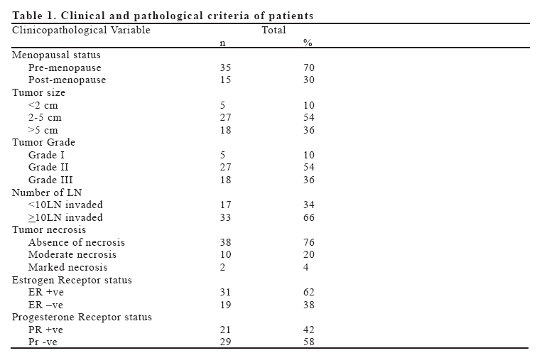
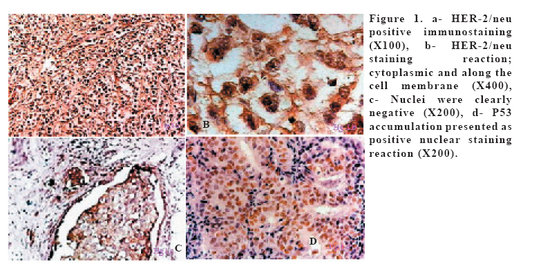
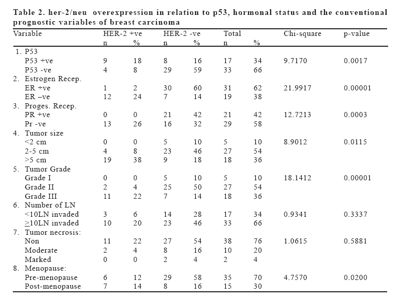
European Journal of General Medicine, Vol. 4, No. 2, 2007, pp. 73-79 THE ASSOCIATION OF HER-2/NEU OVER-EXPRESSION IN RELATION TO P53 NUCLEAR ACCUMULATION, HORMONAL RECCEPTOR STATUS AND COMMON CLINICO-PATHOLOGICAL PROGNOSTIC PARAMETERS IN A SERIES OF EGYPTIAN WOMEN WITH INVASIVE DUCTAL CARCINOMA Mona M Rashed1, Noha M Ragab2, Manal K Galal1 General Organization for Teaching Hospitals & Institutes1, Medical Research Institute, Alexandria University2, Cairo, Egypt Correspondence: Dr. Mona M. Rashed; MD, 47 Mufak Hatata St. Camp Caesar Sq. (21525) Alexandria; Egypt. E-mail: mona_rashed@ems.org.eg Code Number: gm07018 Aim: HER2/neu protein has garnered a great deal of interest in the popular media. However, it has long been known among pathologists and oncologists for its potential role as a tumor and prognostic marker. This protein exists on the surface of epithelial cells and functions in the normal cell as a receptor for a cellular growth factor. Aim of the study: is to determine the association of HER-2/neu and p53 as well as hormonal receptor status with common pathologic parameters in invasive ductal carcinoma of the breast. Key words: Immunohistochemistry, Her-2/neu, p53, hormone receptors, clinico-pathological parameters, breast cancer INTRODUCTION Breast cancer is a heterogeneous disease with variable biological and clinical characteristics. The racial influence in invasive breast cancer in terms of age at presentation, clinico-pathological features, and outcome of treatment has been widely reported. It has been established that breast cancer in many Asian and African countries tend to affect younger females, present in advanced stage with poorer prognostic features, and has a worse outcome when compared to their counterparts in the Western countries. There is no doubt that the lack of early detection and awareness programs contribute to advanced presentation, however, the biological aggressiveness in terms of poor differentiation, lack of steroid receptor expression, and tendency to affect younger females remain unexplained (1). Prognostic factors in breast cancer have exploded over the past several years. Pathologists have played a major role in identifying different histological and immunohistochemical markers that have a direct bearing on both the behavior and treatment of breast cancer (2). Besides several prognostic factors like tumor size, histological grade, steroid hormone receptor status, DNA ploidy and lymph node status which are significant in the management of breast cancer. HER-2/neu, the human epidermal growth factor receptor 2 status might also serve as an additional parameter. HER-2/neu proto-oncogene is involved in the regulation of normal cell growth and division and is expressed at low levels in many normal epithelial cells (3). HER-2/neu proto-oncogene encodes an 185KD glycoprotein with tyrosine kinase activity. Over-expression of this gene either due to gene amplification and/or increased transcription has been observed in a variety of cancers and has been associated with a more aggressive disease and a poor clinical prognosis in 20-30% of patients with breast cancer (4). Over-expression of the proto-oncogene HER2/neu in breast cancer and other tumors appears to correlates with poor survival and may be partially responsible for cellular progression of the neoplastic phenotype. Moreover, HER-2/neu may potentiate tumorgenesis by inducing tumor cell resistance to host defense mechanisms (5) as it induces resistance to tumor necrosis factor (TNF), which causes cancer cells to escape from host immune defenses (6) and appears to result in reduced sensitivity to immune effectors killing (7). The aim of this study is to determine the association of HER-2/neu over-expression in relation to p53 nuclear accumulation, hormonal receptor status as well as conventional common pathologic parameters in a series of Egyptian women with invasive ductal carcinoma of breast. MATERIAL AND METHODS Fifty tumor samples from patients diagnosed as infiltrating ductal carcinoma were included in the present study , they were collected from the pathology department, Damanhour Medical Institute; the General Organization for teaching Hospitals and Institutes. All patients had operable tumors with no evidences of distance dissemination at the time of diagnosis and underwent modified radical mastectomy as well as axillary lymph nodes dissection and removal. All tissues were fixed in 4% buffered neutral formaldehyde at room temperature for no more than 24 hrs. Tumor tissues as well as dissected lymph nodes were examined microscopically. Histology was assessed for tumor subtypes, invasiveness, nuclear and histological grades and lymph node status. Carcinomas were classified on the basis of the H&E stained slides; all cases were invasive ductal carcinoma not otherwise specified (NOS) type and they were graded according to Scarff Bloom-Richardson system of the World Health Organization (WHO) classification (8). Immunohistochemical staining One 4-μm section from each submitted paraffin block was first stained with haematoxylin and eosin in order to verify that an adequate number of invasive ductal carcinoma cells were present and that the quality of fixation was sufficient for immunohistochemical analysis. Serial sections (4-μm) were prepared from selected blocks and float-mounted on adhesive coated glass slides for immunostaining. Known positive control sections were included in each run to ensure proper immunostaining. Sections were processed using a hot citrate buffer for antigen retrieval (2). The status of HER2 (DA 485 dil: 1:1500) and p53 (DO7 dil: 1:200), estrogen (6F11 dil.: 1:40) and progesterone receptors (1α6 dil.: 1:30), were determined by immunohistochemistry on paraffin-embedded sections 4 μm thick. Immunostaining was performed with a Nexus automated immunostainer (Ventana, Illkirch, France). Sections were scored semi-quantitatively by light microscopy by three pathologists. For estrogen and progesterone receptors and p53, a threshold of 10% of stained nuclei was considered positive. Also for HER-2/neu, over-expression corresponded to more than 10% of cells showing complete membrane staining with high intensity. Statistical analysis The HER-2/neu over-expression in breast cancer tissues was studied statistically in relation to prognostic factors including: p53 nuclear accumulation, hormonal receptor status as well as conventional common clinico-pathologic parameters. To evaluate the statistical significant in the present study, Chi-square value with one degree of freedom was applied when appropriate; in the present study a p-value of <0.05 was considered significant. RESULTS The 50 cases included in the present study showed an age incidence which ranged from 26ys-59ys. Thirty five cases, 70% were pre-menopausal and the other fifteen cases (30%) were post-menopausal. Five cases (10%) had a tumor size <2cm, twenty seven cases (54%) had a tumor size 2-5 cm, while eighteen case (36%) had a tumor size >5cm. All cases presented with lymph node metastasis; seventeen cases (34%) had <10 LN invasion and thirty-three cases (66%) had >10 LN invasion. Tumor grades; 5 cases (10%) were grade I, 27 cases (54%) were grade II and 18 cases (36%) were grade III. Tumor necrosis was marked in 2 cases (4%), moderate in 10 cases (20%) and absent in 38 cases (76%) ( Table:1). HER-2 over-expression HER-2/neu positive immunostaining was observed in 13 cases (26%) the staining reaction was cytoplasmic and along the cell membrane; nuclei were clearly negative (Figure 1: a, b & c) (Table:2). Her-2/neu over-expression in relation to Hormonal status ER nuclear immunoreactivity was positive in 31 cases (62%) , where 30 cases (60%) of these were HER-2 negative on the other side 19 cases (38%) were ER negative, 12 cases (24%) out of them were positive for HER-2 cellular accumulation. Statistically there is a highly significant association between HER-2 cellular accumulations and loss of ER nuclear positive immunostaining (p=0.0001). As regard the PR immune-staining 21 cases (42%) were PR positive, all of which were negative for HER-2 immunostaining. Statistically there was also significant association between HER-2 cellular accumulation and PR negative immunostaining (p=0.0003) (Table:2). HER-2/neu over expression in relation to P53 Nuclear accumulation of p53 (Figure 1: d) was recorded in 17 cases (34%); there was a significant association between HER-2 over-expression and the accumulation of nuclear p53 (p=0.0017) (Table:2). HER-2/neu over-expression in relation to Tumor size Most of the HER-2 positive immunostaining cases had a tumor diameter >5cm. Statistically there is a significant association between HER-2 positive immunostaining and the increased size of tumor (p=0.0115) (Table:2). HER-2/neu over-expression in relation to Tumor Grade There was a highly significant association between HER-2 accumulation and the high grade tumors (p=0.00001) (Table:2). DISCUSSION The aggressive biological behavior of invasive and metastatic cancer is considered to be the most insidious and life threatening aspect for breast cancer patients. It is mostly the result of changes in many molecular characteristics of tumor cells, including alterations in mechanisms controlling adhesion, growth and proliferation (9). The biology of breast cancer remains poorly understood as knowledge about individual prognostic factors provides limited information (10). A wide variety of morphology-based and molecular-based breast cancer prognostic factors and tumor markers have been studied. An expanded understanding of the biology of breast cancer has led to the identification of the HER-2/neu receptor as an important growth factor (12). This receptor possesses intrinsic tyrosine kinase activity and has been associated with aggressive biological behavior and poor clinical outcome. Following the original study by Salmon and coworkers in 1987 (13) many investigators have considered the prognostic potential of the HER-2/neu gene and protein in breast cancer (14). The HER-2/neu proto-oncogene encodes a growth factor receptor that is over expressed in 20-30% of metastatic breast cancers. This over expression is associated with decreased survival and decreased relapse-free periods. HER-2/neu gene amplification is also a predictive marker of responsiveness to selected forms of therapy. It is an important gene in breast cancer because patients with HER-2/neu amplification generally have a poor prognosis. Clinical studies have demonstrated that alterations in HER-2/neu predict poor prognosis for breast cancer and are associated with features of tumor aggressiveness, such as absence of estrogen and progesterone receptors, high rate of cellular proliferation, advanced tumor stage, large tumor size, and young age at diagnosis (15). Wild-type p53 plays two major roles in cell function: first, it regulates the checkpoints G1 to S and G2 to M of the cell cycle, through regulation of transcription of p21 and other genes; second, it induces apoptosis after genotoxic damage. Simultaneous over-expression of c-erbB-2 and p53 has been reported to be prognostically unfavorable in breast cancer (16); the development of p53 abnormalities might accompany or precede the development of Her-2/neu over-expression (17). Tumors with both HER-2 over-expression and p53 protein accumulation were reported in several studies (18 &19) and patients with such tumors were found to have poor prognosis. In the present study findings reported indicated that both HER2 over-expression and p53 protein are significantly associated. On the other hand, some studies have shown a better prognosis in patients with breast cancers with HER-2/neu over expression and p53 protein accumulation as they suggested that differences may reflect the effect of various therapeutic regimens (2). HER-2/neu amplification and ER/PR alterations are early events in breast carcinogenesis (14). An inverse association had been found between Her-2/neu amplification/ over-expression and the presence of receptors for steroid hormones estrogen and progesterone in both clinical correlative studies and experimental models; thus the higher the level of Her-2/neu over expression the lower the corresponding ER level (20). Our data also demonstrated an inverse correlation between Her-2/neu over expression and ER status, also there was an inverse correlation between Her-2/neu and PR status that most likely occur because suppression of ER expression leads to reduced expression of PR. HER-2 status has been used as a marker for breast cancer prognosis in addition to the conventional classic factors as lymph node status, tumor size, type and grade (21). In the present study Her-2/neu over expression was significantly associated with the tumor size (p=0.0115) and tumor grade (p=0.00001). Others studies data indicate the association of HER-2/neu positively with large tumors (22 &23). Unexpected finding; there was no significant association between the Her-2/neu over expression and the number of invaded lymph nodes; in the present study all cases had lymph nodes involvements. Others studies showed a direct association between HER-2 and metastatic involvement of lymph nodes (24&25) meanwhile; Sutterlin et al. reported that there was no significant relation between HER-2 over expression and lymph node involvement (26). Korkolis et al. added in their study that deregulation of HER-2 oncogene might characterize a subgroup of node negative patients with poor prognosis who could benefit from an aggressive adjuvant therapy (27). Cell death relates to the inactivation of HER-2/neu receptor in breast cancer (28). Others study mentioned a significant relation between HER-2/neu down regulation and tumor necrosis (29 &30). Another unexpected finding in the present study; the absence of significant association between the Her-2/neu over expression and the grade of tumor necrosis. Zhou et al. suggested that HER-2/neu constitutively activates the anti apoptotic cascade to confer resistance to TNF (Tumor necrosis factor) on cancer cells and reduce host defences against neoplasia (6). Egeblad & Jattela study’s data didn’t support the role of Erb-B receptors in regulation of cell death (31). Finally, there was no significant association between Her-2/neu over expression and menopausal status. Same finding was reported in Adebamowo and coworkers study (32). In Ray et al study; HER-2/neu over expression was significantly higher among post-menopausal in comparison with pre-menopausal women(24). In conclusion this study indicated that Her-2/neu may be a powerful predictor of poor prognosis as it’s over expression was strongly correlated with p53 nuclear accumulation, absence of hormonal receptors immunoreactivity and also unfavorable clinico-pathological variables as tumor size and tumor grade of breast carcinoma. Meanwhile; there was no significant association between Her-2/neu over-expression and tumor necrosis, number of invaded lymph nodes and the menopausal status. REFERENCES
Copyright 2007 - Medical Investigations Society The following images related to this document are available:Photo images[gm07018t1.jpg] [gm07018f1.jpg] [gm07018t2.jpg] |
| |||||||||
