 |
| About Bioline | All Journals | Testimonials | Membership | News |
|
||||||
|
||||||
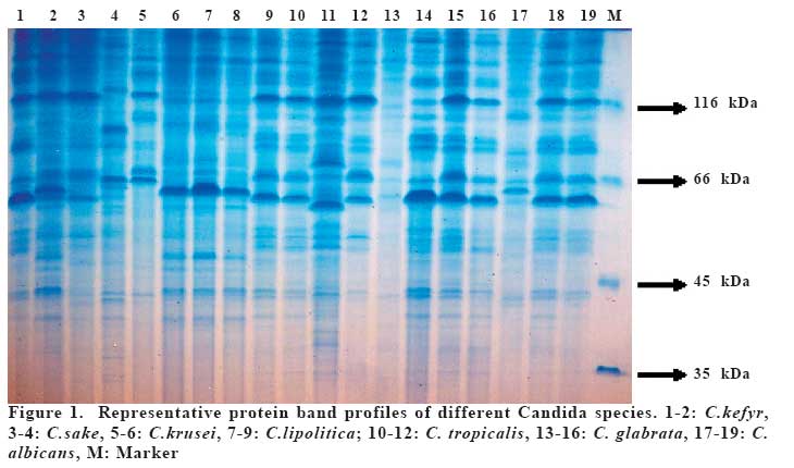
European Journal of General Medicine, Vol. 4, No. 3, 2007, pp. 100-106 NUMERICAL ANALYSIS OF CANDIDA SPECIES FROM URINER SYSTEM INFECTIONS BASED ON SDS-PAGE AND DETECTION OF ANTIFUNGAL RESISTANCE Nizami Duran1, Fatma Öztürk2, Leyla Açık2, Özkan Aslantaş3, Gönül Aslan4 Mustafa Kemal University, Faculty of Medicine, Department of Microbiology and Clinical
Microbiology1, Hatay, Gazi University, Faculty of Arts and Sciences, Department of Biology2,
Mustafa Kemal University, Faculty of Veterinary Medicine, Department of Microbiology3,
Hatay, Mersin University, Faculty of Medicine, Department of Microbiology and Clinical Microbiology4, Mersin, Turkey Code Number: gm07025 Aim: The aim of the present study was to evaluate the protein patterns and numerical analysis of Candida species isolates from urinARY system infections by sodium dodecyl sulphate polyacrylamide gel electrophoresis (SDS-PAGE), and to detect antifungal susceptibility.
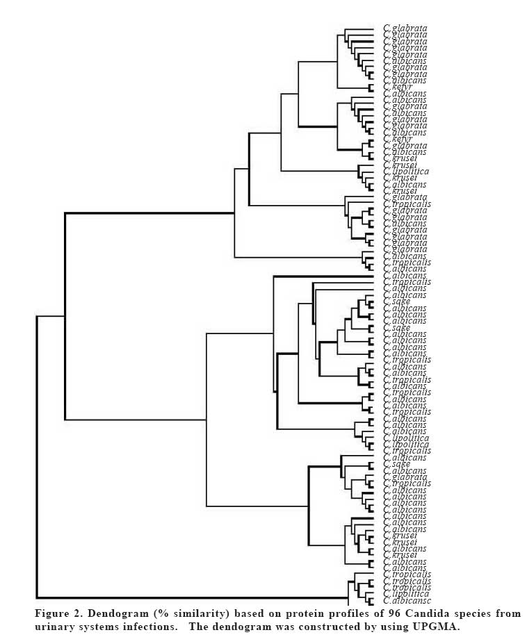
Key words: Numerical Analysis, SDS-PAGE, Candida spp., uriner system infection, antifungal resistance. INTRODUCTION The Fungi, especially yeasts belonging to the genus Candida are potentially pathogenic agents. Yeasts are the most common fungi isolated from human patients. Candida strains are opportunistic pathogenic fungus in humans which can cause either septicaemic or mucosal infections (1). Persons carry the yeast Candida albicans and other Candida species as part of their commensal microflora. However, in hosts predisposed to candidiasis, such as AIDS, diabetes, organ transplant, tumors and others, these yeasts may act as pathogens (2). Commensal Candida species inhabiting the oral cavity, vaginal canal, and gastrointestinal tract of host may begin the infectious process (3-5). Their incidence has greatly increased over the past several decades with the introduction of broad-spectrum antibiotics, immunosuppressive corticosteroids, and antitumor agents as well as an increasing number of AIDS patients (6, 7). For instance, C. albicans is the second cause of nosocomial urinary tract infections in the intensive care unit according to the National Nosocomial Infection Surveillance System reports (8). It is very important to investigate the origin of the Candida isolates that cause nosocomial infections because of high mortality and morbidity of Candida infections (8, 9). For this purpose, several methods have been developed for the characterization or typing of Candida species including morphotyping (10), resistogram typing (11), karyotyping (12), restriction endonuclease analysis of genomic DNA (13). Sodium dodecyl sulphatepolyacrylamide gel electrophoresis (SDS-PAGE) has been employed to analyse whole-cell proteins of candida species. In addition, this technique has been applied to taxonomic studies of Candida species and molecular systematics combined with computerized analysis of proteins (14-18). The purpose of this study was to compare the electrophoretic profiles of different Candida species isolated from urine specimens through the whole cell proteins, and to evaluate their implications for taxonomic purposes by computer assisted numerical analysis. Also, we aimed to investigate the relation between protein profiles and resistance patterns. MATERIALS AND METHODS Candida stains Previously isolated and identified 96 Candida spp. (46 C. albicans, 18 C. glabrata, 14 C. tropicalis,8 C. krusei,6 C. parapsilosis,3 C. dubliensis and 1 C. kefyr) urine were used in this study. Patients whose urine cultures yielded 105 cfu ml-1 or more were selected. Identification of Candida isolates was performed by investigating colony morphology, germ tube formation, microscopic morphology on corn meal agar (Oxoid, UK) with Tween 80 and confirmed by API 32-C System Biomerieux yeast identification programme (Bio-Merieux, France) (19). Whole-cell protein extraction Whole cell proteins of samples were extracted according to modified Kishore method (20). Briefly, all strains were activated in 5 mL YPD medium (2% glucose, 2% peptone, 1% yeast extract ) in a shaker table under 150 rpm, at 30oC, overnight. All cultures were transferred to 50 ml culture and further growed for 24 h at 30oC, in a shaker table under 150 rpm of agitation. After growth, cells were harvested by centrifugation at 4000g for 5 min and pellets were washed twice with distilled water. Two ml of the phosphate buffer (K2HPO4and KH2PO4pH7.0) was added to pellet, and homogenized with sonicater. Seventyfive µl of homogenate and 25 µl of sample buffer were combined and heated in a boiling water bath for 10 min. SDS-PAGE analysis SDS-PAGE was performed according to Laemmli (21), using 4.5% stacking gel and 12.5% (w/v) separating gels. The gel was run at a constant current of 20 mA through stacking gel and 35 mA through separating gel. Proteins in the gel were stained with Coomassie Brillant Blue (22, 23). Protein standarts used for estimation of molecular weight were: 116 kDa ; β-galaktosidase, 66 kDa ; Bovine serum albumin, 45 kDa ; ovalbumin, 35 kDa ; lactate dehydrogenase, 25 kDa ; restriction endunuclease Bsp981, 18 kDa ; β-lactoglobulin and 14 kDa ; lysozym (MBI Fermentas). Antifungal susceptibility testing The susceptibility of Candida strains against amphotericin B and flucanazole were assessed by minimal inhibitory concentrations (MIC). MICs were determined by broth microdilution method following the procedures recommended by the National Committee for Clinical Laboratory Standards (24). The final inoculum size was prepared in modified RPMI-1640 medium (Sigma) as 1000 cfu/ml. Then cells were placed onto flat bottomed microplates (with 96 well) contained two critical concentrations of each drug. The following breakpoint concentrations ranging from 64-0.125µg/ml were studied. The microtiter plates were incubated at 37°C and read visually after 24 h. The MIC values were recorded as the lowest concentrations of the substances that had no visible turbidity. The strains were classified as susceptible, when no growth was observed at both concentrations of drug, intermediate susceptible, when growth was inhibited only in higher concentration or resistant, when strains grew in both concentrations. Isolates for which MICs were ≥64 µg/ ml were accepted resistant to fluconazole and for which MICs were between 16 and 32 µg/ml were considered as dosedependent susceptible (D-DS). As there is no interpretative breakpoint for amphotericin B according to M27-A document, we determined only MIC values for this antifungal agent (NCCLS, 2002). C. albicans ATCC 90028 and C. krusei ATCC 6258 were used as a quality control. Statistical analysis Presence (1) or absence (0) of specific bands was recorded. Similarity dendrograms were built using the unweighted pair-cluster method with arithmetic averages (UPGMA) with the POPGENE software package, version 1.70. Cluster analysis of whole cell proteins was performed according to the genetic distance method of Nei (25) . RESULTS Total protein analysis Total cell proteins of 96 Candida strains were isolated. SDS-PAGE analysis revealed the presence of approximately 30 distinct protein bands with molecular weights ranging in size from 63 to 120 kDa (Figure 1). Although each species produced a characteristic band pattern, some differences in band patterns were observed within the species. The protein profiles of the isolates on gels were reproducible after four repetitions of each electrophoretic running. The application of UPGMA clustering method allowed to building similarity dendrograms based on genetic distances (Figure 2). All Candida species are divided into two main clusters. Lower groub includes 5 C. albicans,2 C. tropicalis and one C.krusei. Upper group is subdivided into two main groups, first upper group contains five subgroups, include18 C. glabrata,9 C. albicans ,2 C. kefyr,4 C. krusei,3 C. tropicalis. Second group is divided into two groups, first group includes 19 C. albicans,3 C. sake, 5 C. tropicalis,2 C. lipolitica. Second group includes 1 C. glabrata,4 C. tropicalis,3 C. krusei, 10 C. albicans,1 C. lipolitica. Antifungal susceptibility testing Minimal ihnibitor concentration (MIC) of Amphotericin B was not higher for C. albicans and non-albicans strains. All Candida strains were sensitive to Amphotericin B in MIC values within range of 0.25-1 µg/ml (1>MIC≥0.25). However, 2 of C.glabrata and 3 of C. krusei strains were resistant to flucanozole. Out of 14 C. tropicalis, flucanozol resistance was not found, 6 of 9 C.krusei strains were found D-DS, and 3 were found resistant. MIC values of flucanzol resistance for 6 C.krusei strains having D-DS were detected to be 0.5-16 µg/ml. Resistance against either Amphotericin B or flucanozle were not detected in other Candida species (C. parapsilosis, C. dubliensis ve C. kefyr). DISCUSSION Candida infections are the most common opportunistic infection among the immuncompromised patients, such as infected-HIV patients, or those living together in the same environment in hospital wards, inter-human transmission of pathogenic fungi is likely to occur frequently. C. albicans and the non-albicans species of Candida are the major agents of candiduria and are emergent pathogens of the urinary tract in critically ill patients. Urinary tract infection caused by Candida strains are increasing nosocomial problem (26, 27). However, it is a rare event and has only recently been demonstrated by molecular typing methods for nosocomial Candida infections in patients at risk for candidosis (28, 29). The analysis of electrophoretic profiles of proteins has allowed the identification, classification of numerous strains, species and genera of yeasts in taxonomic and epidemiological studies (14-18). In the present study, 96 Candida strains from different patients with urinary tract infection were analyzed by SDS-PAGE and numerical analysis. The reproducibility of the electrophoretic protein profiles on different slab gels, evaluated by the inclusion of molecular weight markers and protein extracts of candida strains. The similarity of the electrophoretic whole cell protein patterns among Candida strains samples observed in UPGMA dendrograms showed values between 0 % and 99 %. The data obtained from grouping of Candida strains based on their electrophoretic profiles showed high level of agreement with the inter-specific classification established by conventional methods. Moreover, the isolates of each species showed identical or very similar profiles when compared (Figure 1). This fact suggests that these protein profiles obtained by SDS-PAGE are relatively stable taxonomic characteristics. This method shows good reproducibility and allows collection of useful information for numerical analysis. This methodology brings relevant information in systematic evaluation of related species. This study showed that the SDS-PAGE technique has proved to be a useful method for systematic or epidemiological purposes. Azole-antifungals is the largest and most widely used class of antifungal agents. Recently, high antifungal (azole) resistance in non-albicans strains especially C. glabrata and C. krusei have been reported (6, 30). In this study, we detected D-DS resistance in 6 of 9 C. krusei strains. Amphotericin B resistance among Candida strains except C. lusitaniae have been reported to be low (31). Also, in our study, we could not detect amphotericin B resistance among Candida species isolated from immuncompetent persons. But, it has been shown that amphotericin B resistance could be high in immunsupressed patiens such as neutropenic patients, and may pose serious problem (32). Antifungal resistance of Candida spp. has been reported to be related with changes or differences occured at cell wall and plasma membrane (34). Alhough it was aimed to investigate relationship between protein profiles and antifungal resistance, we could not detect any relation between antifungal resistance and protein profiles. In conclusion, differentiation and numerical analysis of Candida species based on SDS-PAGE may provide preliminary criteria for taxonomic and epidemiological studies of such microorganisms. Besides this, the similarity among Candida strains isolated from the patients with urinary tract infections was observed in our hospital. REFERENCES
Copyright 2007 - Medical Investigations Society The following images related to this document are available:Photo images[gm07025f2.jpg] [gm07025f1.jpg] |
| |||||||||
