 |
| About Bioline | All Journals | Testimonials | Membership | News |
|
||||||
|
||||||
European Journal of General Medicine, Vol. 5, No. 1, 2008, pp. 21-26 Histological Investigation Of Testicular And Accessory Sex Glands In Ram Lambs Immunized Against Recombinant Gnrh Fusion Proteins Öner Odabaş1, Mehmet Kanter2 Yuzuncu Yıl University, Faculty of Medicine, Department of Urology1 , Van, Trakya University, Faculty of Medicine, Department of Histology and Embryology2, Edirne, Turkey Code Number: gm08004 ABSTRACT Aim: The aim of the study was to investigate the histological appearance of testis, epididymis, prostate and seminal vesicles of ram lambs immunized against gonadotrophin releasing hormone (GnRH).
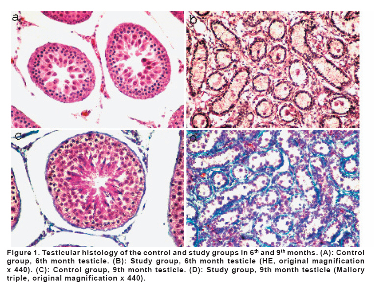
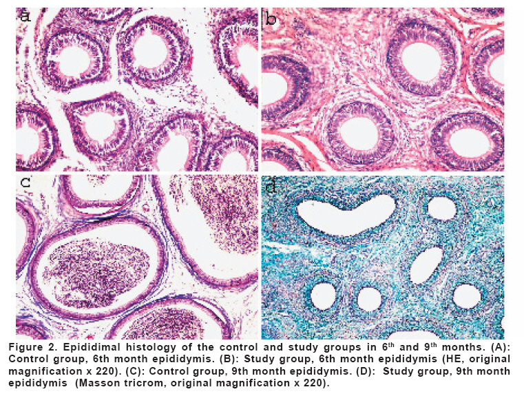
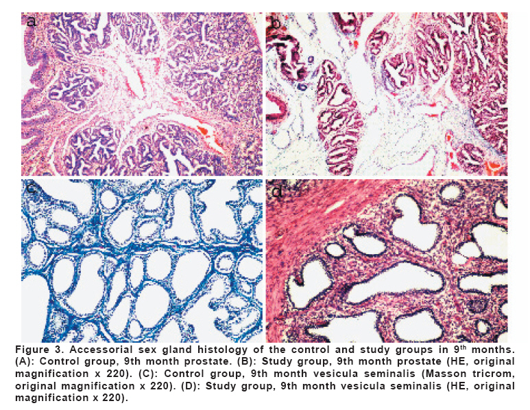
Key words: Recombinant GnRH fusion protein, testis, epididymis, prostate, vesicula seminalis. INTRODUCTION Gonadotrophin releasing hormone (GnRH) is the primary hormone controlling reproduction in mammals. Giving GnRH agonist and antagonist or immune neutralization against GnRH decreased gonadotroph hormone concentration and consequently blocked testicular growth and sexual activity (1). A sterilization vaccine containing the GnRH molecule and a binding carrier protein has been produced by recombinant technology. Several veterinary studies have been carried out with this recombinant fusion protein vaccine. It has been considered as an alternative sterilization agent to surgical castration in farm animals (2, 3). This vaccine has also been used in humans for several aims, such as lengthening lactational amenorrhea as a contraception method in postpartum women and treatment of prostate cancer (4). The aim of the study was to evaluate the effectiveness of a recombinant GnRH fusion protein vaccine in suppressing testicular and accessory sex gland (epididymis, prostate and seminal vesicles) development in young ram lambs. MATERIAL AND METHODS This study was performed on 14 male lambs, 7 in the study group and 7 in the control group. Another 5 lambs were included from the study after biopsy samples were taken from their testes at age 10 weeks to determine testicular histology at the start of the study. The animals were fed a diet of clover hay and cracked barley with a vitamin-mineral supplement. Seven lambs aged 10 weeks were immunized against GnRH using recombinant fusion proteins (ovalbumin luteinizing hormone releasing hormone (LHRH-7) and thioredoxin LHRH-7). Immunization was repeated twice monthly (booster immunizations) in the study group until slaughter at 9 months. The control group was not immunized. Testes and epididymis biopsies of both groups were taken after 6 months. Ovalbumin LHRH-7 and thioredoxin LHRH-7 fusion proteins were prepared by recombinant DNA technology as described previously (5, 6). A total of 1 mg of the mixture, including equal amounts of the two proteins, was emulsified in 6 M urea then injected into the study group animals. The protein mixture was given with 0.5 ml Freund’s modified complete adjuvant at first immunization and with Freund’s incomplete adjuvant at the booster immunizations. Injections were made subcutaneously into the lower parts of fore legs in equal amounts at each immunization. All the animals were slaughtered at 9 months of age, then testis, epididymis, prostate and seminal vesicles were extracted for histological investigation. Biopsy specimens were put in 10 percent neutral buffered formalin solution. Tissue samples were then processed for histological examination. Six micrometer-thick sections from paraffin-embedded samples were stained by Mallory triple (Figure 1C, D), Masson tricrom (Figure 2C, D, Figure 3C) and hematoxylin and eosin (H&E) (Figure 1A, B, Figure 2A, B, Figure 3A, B, D) techniques. Stained preparations were investigated with a Nikon optiphot-2 microscope. RESULTS In the control group, 10-week-old lamb testes had the appearance of infantile testes: their tubular diameter was very small and spermatogenesis had not started. In testes at 6 months, spermatogenesis had started but spermatozoa were immature. At 9 months testes appeared completely mature, with spermatozoa visible in the tubule lumen. Histopathology of the testes in the study group at 6 and 9 months was similar, in that tubule diameter showed significant regression (Figure 1B, D, respectively). No developing spermatogenic cells could be seen in the tubules and cellular density had increased in the intratubular space. At 6 and 9 months, testes in the control group included cells in the development phases of spermatogenesis (Figure 1A, B, respectively) whereas in the study group of the same ages, Sertoli cells lined the base of the tubules. Spermatogonia were rarely seen in the tubules. The epididymis of the control and study groups at the 6th month did not differ significantly, except for an increase in intertubular connective tissues (Figure 2A, B, respectively). In the control group epididymis at 9 months, tubule volume was greater and sperm cells had formed a sperm ball in the tubule compared with the control group at 6 months (Figure 2C). In the study group at 9 months tubule volume was decreased and intertubular connective tissues were increased in comparison with the control group at 9 months. Leydig cells lying in between the seminiferous tubules were virtually non-existent and rarely in small clusters, leaving only a few irregular polyhedral cells in rows together with blood vessels. No spermatozoa was seen in the epididymal tubule, and epithelial cells lining the tubule showed hyperplasia (Figure 2D). The two groups’ prostates differed slightly but not significantly at 9 months: asiner epithelial cells of the control group had a slightly richer cytoplasm than those of the study group and there was mild stromal increase in the study group (Figure 3A, B). At 9 months the seminal vesicles of the study group had an excessive stromal increase compared with the control group (Figure 3C, D). DISCUSSION Active immunization against GnRH and its analogues has a significant effect on fertility in males and females of different species (7-10). Immune neutralization of GnRH decreased gonadotroph hormones and sex steroids, with eventual gonadal atrophy. Decreasing the antibody titration restored fertility, that is, the effects of immunization are reversible (11,12). Histology is an important way of studying the effects of GnRH fusion protein vaccine on the reproductive system. We observed many changes in histological preparations of the study group compared with the control group. Testosterone level, sperm count, sexual activity and fertility of breeding animals have been used as indirect indicators of the effects of GnRH vaccine on testes (13-15). The most significant changes reported as a result of GnRH vaccine have been in the testis (9, 10, 16). Ferro et al. (16) immunized rats with GnRH vaccine and observed testicular atrophy and azoospermia in seminiferous tubules by light microscope. Neutralization of GnRH by active immunization not only suppressed testicular development and testosterone secretion but also reduced spermatogenesis. This is consistent with the anti-gonadal effect of the anti- GnRH vaccine in other species (17,18) and indicates that the fertility of immunized rams was probably markedly reduced. Several studies (19) have examined the effect of neutralization of GnRH on the gonadal function of ram lambs actively immunized against GnRH. Histologic examination of tissue samples collected at slaughter indicated that seminiferous tubule diameter was reduced in testes of Freund’s complete adjuvant lambs, relative to tubule diameter in untreated lambs. Atrophy of seminiferous tubules was also noted in 82, 25, and 0% of testes collected from Freund’s complete adjuvant, another oil-based adjuvant, and untreated lambs, respectively. Testes collected from Freund’s complete adjuvant lambs were completely devoid of spermatozoa. In contrast, spermatozoa were evident in 50 and 69% of the testes of lambs in the another oil-based adjuvant and untreated groups, respectively. These investigators found clear evidence of secondary infertility, as we did, but, in contrast to our results, they observed rare spermatozoa in the caput epididymis. Likewise in our study, Ferro et al. (16) indicated that the histological assessment showed drastic changes in testicular morphology, as well as atrophy to the seminiferous tubules and a total depression in spermatogenesis in the GnRH-I immunised animals. Marked atrophy of the testis leading to testicular failure was characterised by azoospermia of the seminiferous tubules. The spermatogenic cells of the tubules appeared to become degenerated and were continuously cast off. The inner wall of the tubules contained only a small number of spermatogenic cells (spermatogonium and spermatocytes) and Sertoli cells, with no visible spermatozoa under light microscopy (16). Leydig cells lying in between the seminiferous tubules were virtually non-existent and rarely in small clusters, leaving only a few irregular polyhedral cells in rows together with blood vessels. The effects of LHRH fusion protein on ram epididymis and seminal vesicles in our study were similar to the results of studies done on other species. Ferro et al. (16) found tubular folding and hyperplasia of tubule epithelia in all parts of the rat epididymis immunized with GnRH vaccine. Pseudo stratified columnar epithelia cells of ductuli efferentes changed into tall stratified columnar cells. Sperm concentration in ductuli efferentes declined and no sperm were seen in the cauda epididymis (16). Our epididymal histological findings were similar to those of Ferro’s study except for the existence of sperm cells. Giri et al. (20) injected Bonnet monkeys with LHRH vaccine, then investigated sex glands histologically. They showed that LHRH vaccine caused flattening of the glandular epithelia of monkey seminal vesicles and an increase at stromal tissue. These are similar to our findings but their findings on prostate tissue differ from ours. We observed no evident effect of LHRH fusion protein on the prostate, whereas these authors showed a weakness of tall columnar secretary epithelia of the prostate and an increase in stromal tissue proportion to secretary epithelia. In their study the prostate was the most affected of the sex glands. The differences between the findings in the studies reveal that the effect of LHRH vaccine might differ among species. Acknowledgments We thank Dr. Hasan Ulker because of his kind assistance to practice on rams and to acquire the recombinant GnRH fusion proteins. REFERENCES
Copyright 2008 - Medical Investigations Society The following images related to this document are available:Photo images[gm08004f3.jpg] [gm08004f2.jpg] [gm08004f1.jpg] |
| |||||||||
