 |
| About Bioline | All Journals | Testimonials | Membership | News |
|
||||||
|
||||||
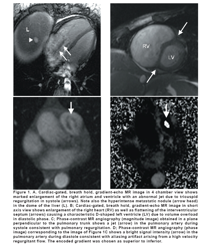
European Journal of General Medicine, Vol. 5, No. 1, 2008, pp. 54-56 Magnetic Resonance Imaging Diagnosis Of Carcinoid Heart Disease Orkun Baloğlu1 , Servet Tatlı2* Hacettepe University, School of Medicine1, Ankara, Turkey, Brigham and Women’s Hospital, Harvard Medical School, Department of Radiology2, Boston, USA Code Number: gm08011 ABSTRACT Carcinoid heart disease is a rare complication of metastatic carcinoid. We present a 46-year-old woman who had metastatic carcinoid and progressive right heart failure. Cardiac MR imaging was helpful in the detection of the extent of the cardiac disease. Key words: MRI, carcinoid tumor, carcinoid heart disease INTRODUCTION Carcinoid heart disease is an uncommon complication of the carcinoid syndrome that often leads to heart failure (1). Histologically, it is characterized by pathognomonic plaque-like intracardiac deposits of fibrous tissue with retraction and fixation of valvular cusps (2). Echocardiography may be limited to demonstrate the extent of the cardiac involvement. MR imaging may be helpful for the demonstration of the extent of the disease in this is rare clinical condition. We describe the magnetic resonance (MR) imaging findings of heart involvement in a patient with metastatic carcinoid. CASE A 46-year-old woman presented with bilateral progressive pedal edema, facial flushing episodes and severe diarrhea. Urine 5-hydroxyindoleacetic acid level was markedly elevated. Abdominal CT examination revealed innumerable liver metastases without a primary site. Liver biopsy was performed and pathologic evaluation revealed the diagnosis of carcinoid tumor. Her echocardiography showed thickened, retracted tricuspid valve with opposition of the leaflets and moderately enlarged right atrium and ventricle consistent with carcinoid disease of the heart. The left heart chambers were unremarkable. The pulmonary valve could not be evaluated. Upon progression of her symptoms, obtained MR imaging to evaluate the extent of the disease demonstrated significantly enlarged right ventricle (Figure 1 A,B) and atrium with evidence of volume overload that manifested by presence of D-shaped left ventricle seen in diastole only (Figure 1B). On gradient echo cine images, the right ventricle function was preserved. The tricuspid valve appeared thickened and retracted with narrow apposition of the leaflets. There was severe retraction as well as severe tricuspid regurgitation (Figure 1A). In addition, the pulmonary valve was also involved with moderate severity of insufficiency (Figure 1 C, D). The aortic and mitral valves were structurally normal with traces of insufficiency. Left ventricle size and function was normal. The patient underwent elective tricuspid and pulmonary valve replacement. Pathologic evaluation of the tricuspid and pulmonary valves revealed chorda tendineae with fibrous endocardial thickening with abundant smooth muscle cells, devoid of elastic fibers consistent with carcinoid plaque. The patient was discharged in a stable condition. DISCUSSION Carcinoid heart disease is an uncommon complication of the carcinoid syndrome that often leads to heart failure (1). Histologically, it is characterized by pathognomonic plaque-like intracardiac deposits of fibrous tissue with retraction and fixation of valvular cusps (2). It typically involves the valves and endocardium of the right side of the heart. Tricuspid regurgitation is found nearly in all patients but tricuspid stenosis and pulmonary regurgitation less commonly occur (2). Echocardiography is the most commonly used diagnostic tool to identify carcinoid heart disease. Cardiac MR imaging adds valuable information when evaluating cardiac structures that might be difficult to analyze on echocardiography (3, 4). MR imaging features of the carcinoid disease of the heart include dilatation of the right atrium and ventricle with flattening of the ventricular septum; thickening of tricuspid valve and its subvalvular apparatus with marked immobility throughout the cardiac cycle leading a fixed open position of the cusps during systole; and a severe tricuspid regurgitation (4). Although its spatial resolution is limited to demonstrate patognomonic valvular deposits, MR imaging has the advantage that valvular dysfunction can be precisely quantified with phase contrast technique. MR imaging can also be useful in determining the involvement of pulmonary valve, which may sometimes be difficult to evaluate on echocardiography (5). REFERENCES
Copyright 2008 - Medical Investigations Society The following images related to this document are available:Photo images[gm08011f1.jpg] |
| |||||||||
