 |
| About Bioline | All Journals | Testimonials | Membership | News |
|
||||||
|
||||||
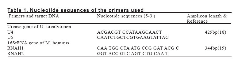
European Journal of General Medicine, Vol. 5, No. 2, 2008, pp. 107-111 Comparison Of Culture With The Polymerase Chain Reaction For Detection Of Gennital Mycoplasma Shahin Najar Peerayeh and Roghayeh Samimi Tarbiat Modares University, School of Medical Sciences, Department of Microbiology Tehran, Iran Code Number: gm08020 Aim: Genital mycoplasmas are known as sexually transmitted agents, causing mainly urethritis, pelvic inflammatory disease, spontaneous abortion, pyelonephritis, infertility, still birth, low birth weight, neonatal meningititis, and neonatal pneumonia. Mycoplasma infections not only jeopardize fertility but also pose a risk for infertility treatment and resulting pregnancies. Diagnosis of genital mycoplasma infections by bacterial conventional methods is very difficult. The aim of this study was to comparison of culture with polymerase chain reaction (PCR) for to determine the prevalence of Ureaplasma urealyticum and Mycoplasma hominis in the endocervical specimens from infertile women.
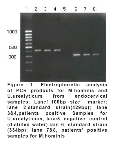
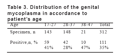
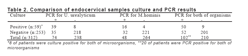
Key words: Ureaplasma urealyticum, Mycoplasma hominis, infertility, PCR INTRODUCTION Mycoplasmas are the smallest cell free-life microorganisms. They can be isolated as commensals or pathogens from plants, insects, animals and humans. Some of them are considered normal flora of the respiratory or genitourinary tract (1). Ureaplasma urealyticum can be found in the cervix or vagina of 40-80% of sexually mature, asymptomatic women and Mycoplasma hominis in 21-53% (2-6). The presence of genital mycoplasmas in a large proportion of healthy women complicate the assessment of the pathogenic roles of these organisms, but several studies have indicated that genital colonization of these mycoplasmas can be associated with an increased risk of developing certain pathogenic conditions and pregnancy abnormalities, e.g., pelvic inflammatory disease, pyelonepheitis, premature rupture of membranes, chorioamnionitis, and preterm labor and birth. In addition, they may be acquired by neonates either in utero or by vertical transmission at birth and can cause pneumonia, pulmonary hypertension, chronic lung disease, and meningitis of the newborn (7-11). In addition, infections with genital mycoplasmas have been linked with infertility (11-16). The main method of detecting mycoplasmas are by culture, but the organisms are difficult to isolate and require special culture media. Polymearase chain reaction (PCR) is revolutionizing the diagnosis of many infectious diseases, particularly those caused by organisms that are difficult to cultivate. In this study we compared culture with PCR for detection of U. urealyticum and M. hominis in endocervical samples from infertile women. MATERIAL AND METHODS Clinical specimens- Between February 2003 and August 2003, endocervical swab samples were taken from a total of three hundred and twelve infertile women, ranging in age from 17 to 45 years, in infertility clinics of Tehran city. From patients a detailed history was obtained, and all complaints concerning the genital tract, such as abortion and cervicitis were recorded. Cultivation of specimens Samples inoculated into the liquid medium (0.6% beef heart, 1% peptone, 0.5% sodium chloride supplemented with 20% horse serum, 10% fresh yeast extract, 0.05% Hcl-cystein, 1% urea, 1.5% arginin, 20000U of penicillin G per ml, and 0.125% phenol red, adjusted to pH 6.5), were incubated at 37C. As soon as the pH of the medium changed, the cultures were centrifuged and the residues were transferred to plates of solid medium (made up of same constituents as liquid medium with the addition of 1.5% agar), and incubated at 37C in an atmosphere of 5% CO2. DNA extraction from specimens DNA was extracted from standard strains (U.urealyticum serotype VIII and M. hominis PG 21 were kindly provided by Statns Serum Institut of Copenhagen, Denmark) and clinical samples as described previously by Cadieux et al (17). Briefly, 1ml of the sample was centrifuged at 12000 ×g for 10 min. The pellet washed in PBS and resuspended in 50µl of distilled water. After boiling for 10 min, an aliquot of 7µl was used directly in PCR experiments. PCR assay PCR reactions were performed with an automated thermalcycler (Eppendorf, USA). Specific oligonucleotide primers were used for U.urealyticum and M. hominis(Table 1). These were chosen in published nucleotide sequences: in the urease gene for U.urealyticum (18) and in the 16S rRNA gene for M. hominis(19). Amplification was performed in 50µl of reaction mixture containing 10µl of 10× PCR buffer, 2.5 mM Mgcl2, 200µM dNTP, 1.25 units of Taq polymerase, 20pmol of each primer and 7µl of sample DNA. The reaction mixtures were placed in thermalcycler. The thermal profile involved an initial denaturation step at 94C for 3min followed by 30 cycles of denaturation at 94C for 1min, primer annealing at 52C for U.urealyticum and 62C for M. hominis for 1min, and primer elongation at 72C for 1min. The cycling was followed by a final extension step at 72C for 10min. Aliquots of amplified samples (10µl) were analyzed by electrophoresis on a 1.5% agarose gel stained with ethidium bromide. Statistical analysis Chi-square (X2) test was used for the generation of P<0.05 values. RESULTS PCRs results Of the 312 patients studied, 102(32/6%) were positive for U.urealyticum, M. hominis or both genital mycoplasmas (Table 1). Of these 102patients, 54 (52.9%) were PCR positive only for U. urealyticum, 28(27.4%) were PCR positive only for M. hominis and 20(19.6%) presented both organisms. A photograph of electrophoresis based on bromide-stained agarose gel for PCR-amplified products from the Mycoplasmas strains is presented in Fig. 1. DNA from the 16S rRNA sequences that is amplified by the PCR primers used in this study shows at 334bp (M. hominis). A 429bp fragment of the urease gene was amplified for identification of U. urealyticum. Mycoplasmas isolation from clinical samples and comparison with PCR results: The prevalence of positive endocervical culture results was 18.9%(59/312). Of the 59 culture positive specimens, U.urealyticum was isolated from 39, M. hominis was isolated from 12 and both organisms were isolated from 8. A comparison of all results from the PCR procedure with those from culture method is shown in Table 1. Of the 312 patient results analyzed, 111(35.5%) were positive by both culture and PCR. 50 specimens were PCR positive as well as culture positive, 72 were positive only by PCR and 9 were positive only by culture. The PCR had a sensitivity of 91.8%(102/111), and culture of 53.1%(59/111). In this population, 37% of the women presented with sings or symptoms, such as vaginal discharge (25%), cervicitis (17%) and abortion (18%). When relating mycoplasmas with those clinical features, these microorganisms were detected in 31% of the patients with vaginal discharge (M. hominis in 4% of women, U. urealyticumin 16% and both microorganisms in 11%). In women with abortion, genital mycoplasmas were identified in 31% (M. hominis in 9%, U. urealyticumin 17% and both microorganisms in 5%). Mycoplasmas also were detected in 43%of patients with cervicitis (M. hominis in 17%, U. urealyticumin 15% and both microorganisms in 11%). The age of the patients from who were PCR positive varied from 17-45 with a mean age of 31 years. Distribution of the genital mycoplasmas in accordance to patient’s age is presented in Table 3. NO significant difference was found between the age of patients whose sample was PCR positive (positive group) and that of the other patients (negative group). There was also no difference regarding the period of infertility, vaginal discharge, and abortion, between the positive and the negative group. DISCUSSION The results indicate that the use of a PCR assay for genital mycoplasma in endocervical samples results in a higher rate of detection of these microorganisms than that observed with standard microbiologic cultures for genital mycoplasmas. Enhanced sensitivity for genital mycoplasmas detection with PCR is consistent with the literature (20-23). This finding is not surprising given the fact that mycoplasmas are liable organisms lacking a cell wall. PCR has an advantage in that it can still detect DNA of dead organisms. Although the combined use of liquid and solid media is thought to be the most sensitive culture method available, it has been shown in other studies (20) to detect only 75-80% of samples infected with Mycoplasmas. If in our study the111 positive specimens detected by both culture and PCR represented all infections, then the 59 positive specimens detected by culture represent only 53% of the infected samples, whereas the PCR positive samples represent 91% of them. The lower level of detection by culture may be attributed at least in part to the generally recognized difficulties of culturing and isolating mycoplasmas. In addition, the difference may be related to possible effects of uncontrolled ambient temperature between collection and the beginning of culturing in the present study or the effects of other organisms or material present in the specimens. Nine patients in our study had a positive culture but a negative PCR assay result. Similar observations have been reported by other investigators (20,22) and have been attributed to degradation of bacterial DNA or inhibitor(s) of the PCR reaction in clinical samples. For example, blood is a poor biologic specimen for PCR-based methods, because hemoglobin can inhibit the PCR reaction. Blood contamination of endocervical specimens is therefore a potential source of false-negative results. It is noteworthy that despite these potential problems the overall sensitivity of PCR for genital mycoplasma in the detection of microbiologically proven infection of the endocervix by mycoplasmas was 91.8%(102/111). In addition to its greater sensitivity and lesser dependence on careful specimen handling between collections and testing, PCR had a further advantage of faster determination. Assay time was reduced from 2 to 5 days for culture to <24 hours for PCR. In this study, 35.5% of 312 infertile women were colonized with genital mycoplasmas as detected by culture and/or PCR. U. urealyticum(26%) has been shown to be more frequently detected in women than M. hominis(15.7%). Other studies using PCR for detecting genital mycoplasmas in endocervical specimens have reported prevalence rate as high as 40 to 80% for U. urealyticum and 20-50% for M. hominis(2,21,22,23,24). Since genital mycoplasmas have been found significantly associated with low socioeconomic background, such as poverty, number of sexual partners, and use of contraceptive drug (2-6), it is not surprising that the rate of genital mycoplasmas was lower in our study. Although differences were not statistically significant, but the isolation rate of genital mycoplasmas were higher in women under 30 years of age, that is consistent with that previously described by others (2,3,5,25). The use of PCR methodology is increasing in clinical microbiology laboratories, being useful for agents that are costly, slow, and/or difficult to cultivate. Mycoplasmas are good candidates for this technology for all of the above reasons. This study, like others (18,21,24,26) has demonstrated that PCR is a more sensitive and reliable means of detecting genital mycoplasmas in the endocervical specimens. Therefore the specificity, the exquisite sensitivity, and the rapidity of PCR make this technique most valuable in the diagnosis of genital mycoplasmas infections. REFERENCES
Copyright 2008 - Medical Investigations Society The following images related to this document are available:Photo images[gm08020t2.jpg] [gm08020t3.jpg] [gm08020t1.jpg] [gm08020f1.jpg] |
| |||||||||
