 |
| About Bioline | All Journals | Testimonials | Membership | News |
|
||||||
|
||||||
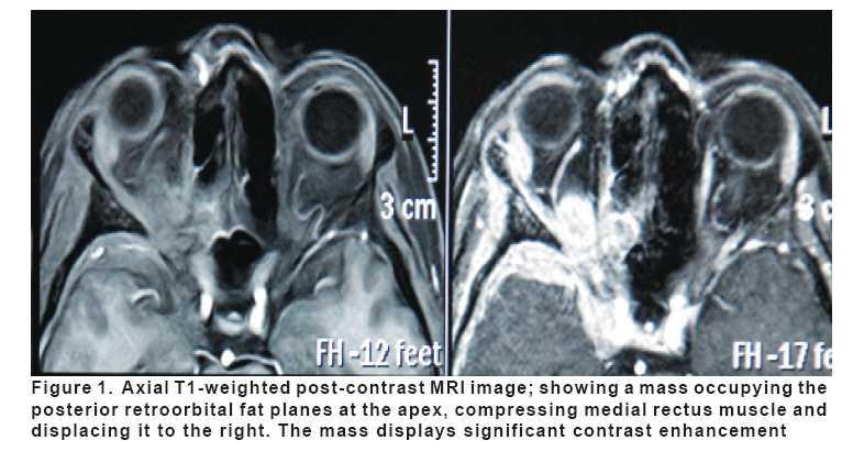
European Journal of General Medicine, Vol. 5, No. 2, 2008, pp. 121-122 Orbita Breast Metastasis Adem Gül1, Adil Kılıç1, Adnan Çınal1, Serhat Avcu2, Tekin Yaşar1 Yüzüncü Yıl University, Faculty of Medicine, Departments of Ophthalmology1 and Radiodiagnostics2, Van, Turkey. Code Number: gm08024 Breast carcinomas are among the most frequent metastatic lesions of the orbit. Diagnosing the disease earlier is of great importance in maximising the life quality of the patient. We report a 35-year-old-female with a retroorbital mass due to the metastases from her left breast invasive ductal carcinoma. Pre-existing malignant diseases should be considered in differential diagnoses of external ophthalmoplegia, central retinal vein occlusion, optic nerve meningioma, and proptosis. The ophthalmologists should be aware that eye, particularly orbita breast metastases may be easily overlooked and a late diagnosis would not work. Key words: Breast cancer, metastasis, orbit. INTRODUCTION Breast carcinomas are among the most frequent metastatic lesions of the orbit (1). The overall prognosis for the patients with orbita breast metastasis still remains poor, despite the treatment, which is usually palliative (1). Diagnosing the orbita breast metastases earlier is of great importance in maximising the life quality. Pre-existing malignant diseases should be kept in mind in differential diagnoses of external ophthalmoplegia, central retinal vein occlusion (CRVO), optic nerve meningioma, and proptosis (2). We report a case with orbita breast metastasis, which is rare and is easily overlooked. CASE A 35-year-old-female presented with right upper ptosis for nearly 1 month. She had mild proptosis in her right eye. The best corrected visual acuity was 0.3 and 1.0, in her right and left eye, respectively. Ocular excursions to all sides were completely restricted in her right eye, whereas they were normal in the fellow eye. Her right pupilla was middilated and direct pupilary reflex was not preserved in her right eye. Fundus examination of her right eye disclosed papilledema. Magnetic resonance imaging revealed a retroorbital mass which was 26x10 mm in size, and which extended mostly at the posterior pole of the optic nerve (Figure). The examinations of her left eye disclosed normal findings. The metastases were consistent with breast origin. She had had a complaint of distortion of her left breast for one year at the time of diagnosis of the breast cancer. Systemic examination revealed invasion of cranium, liver, and lungs by invasive ductal carcinoma of her left breast. Pathological examination of the specimen obtained from her left breast showed grade 3 invasive ductal carcinoma. The lady was administered chemotherapy. DISCUSSION The improved survival of the patients with breast cancer, together with elongation of the mean life time of the population has lead to a higher incidence of the patients with orbital metastates. Moreover, advances in diagnostics caused increases in detecting orbital metastases (1). Ophthalmic manifestations of metastatic breast carcinoma was reported in 5.8% of asymptomatic cases (3). Orbital metastases were reported to occur in 1.45% of the patients in the same study (3). That is to say, not infrequently, metastases of breast carcinoma to the eye are misdiagnosed. The carcinoembryonic antigen was reported as useful in confirmation of the diagnosis of metastatic breast cancer (4). Treatment of orbital metastases include radiotherapy, chemotherapy, hormonal therapy, surgical intervention, or combination of these modalities (1). Bullock and Yanes, analyzing the records of 30 patients with breast cancer, reported that a wide spectrum of ophthalmic manifestations, including cranial nerve involvement, brain involvement with papilledema, Horner’s syndrome, and choroidal and orbital tumors are likely (4). Optic nerve breast metastasis can masquerade as CRVO or optic nerve meningioma(2). Backhouse et al. (2) concluded progressive infiltration of the optic nerve by breast tumor metastases probably enhance CRVO ischemia and resultant rubeotic glaucoma. Metastatic involvement of extraocular muscles by lobular mammary adenocarcinoma, a tumor originating from breast, has been previously reported (5). Metastasis of the breast cancers to eye occurs via haematogenous route. (5,6). We, too, suggested a hematogenic route for the case we present. In our case, decreased visual acuity was the result of optic nerve involvement, restriction of ocular excursions was due to both the mechanical effect of the mass, and involvement of ocular muscles and nerves. Papilledema in our case was due to involvement of optic nerve and metastasis. To conclude, the ophthalmic practitioners should be aware that eye, particularly orbita breast metastases may be easily overlooked and it may be too late to manage an orbital metastasis when a late diagnosis was made. REFERENCES
Copyright 2008 - Medical Investigations Society The following images related to this document are available:Photo images[gm08024f1.jpg] |
| |||||||||
