 |
| About Bioline | All Journals | Testimonials | Membership | News |
|
||||||
|
||||||
European Journal of General Medicine, Vol. 5, No. 3, 2008, pp. 152-156 Formaldehyde-induced Damage In Lungs And Effects Of Caffeic Acid Phenethyl Ester: A Light Microscopic Study Aslı Özdem Türkoğlu1, Mustafa Sarsılmaz1, Neriman Çolakoğlu2, İsmail Zararsız1, Tuncay Kuloğlu2, Hıdır Pekmez3, Ufuk Taş1 Fırat University, Faculty of Medicine, Departments of Anatomy1 and Histology and Embryology2, and Firat University, School of Health Sciences3, Elazig, Turkey Code Number: gm08030 Aim: The aim of this study was to investigate the toxicity of formaldehyde on lung and protective effects of caffeic acid phenethyl ester against these toxic effects.
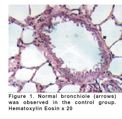
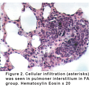
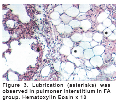
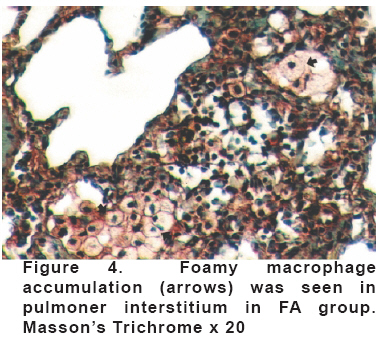
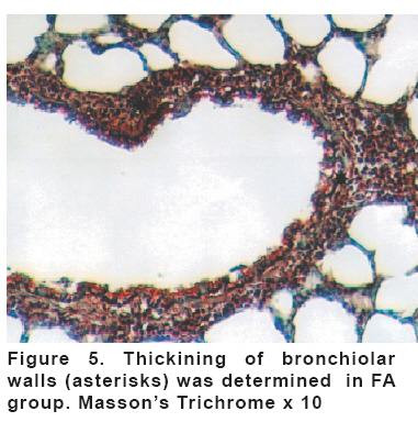
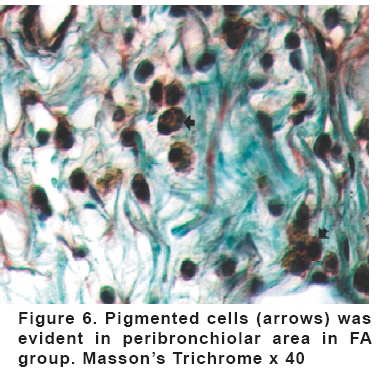
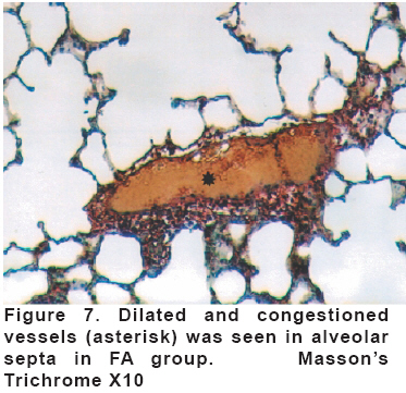
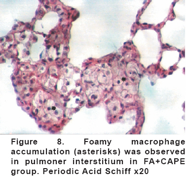
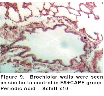
Key words: Formaldehyde, caffeic acid phenethyl ester, lung. INTRODUCTION Formaldehyde is used as a sterilizing agent, disinfectant and preservative in many occupational areas and at home (1). Motor vehicle exhaust, the burning of gas, oil, coal, wood, rubbish and photochemical smog are some environmental sources for formaldehyde. It is colorless gas has a pungent odor and is irritating to the mucous membranes of the nose, throat and eyes (2). The toxicity of formaldehyde is of concern to all who work closely with it. Embalmers, anatomists, technicians and medical, dental or veterinary students are among the people who have high exposure to formaldehyde (3). Formaldehyde poses many potentially detrimental effects to body systems. Many animal studies showed that exposure to formaldehyde cause serious harm on the respiratory system. The primary target for FA-induced toxicity in both rodents and monkeys are the respiratory nasal epithelium (4). Long-term formaldehyde inhalation at a dose of 15 ppm was induced squamous cell carcinomas in the nasal cavities of rats and mice (5). Carpet factory workers who occupationally exposed to formaldehyde were presented loss of respiratory function (6). CAPE is one of the active constituents of the propolis, a substance with a sharp and pleasant odor that is found in the plant extracts accumulated by bees (7). It has been used in the treatment of many diseases by folk medicine (8, 9). CAPE has antioxidant, antibacterial, anti-inflammatory, and antiviral effects. It also presents immune stimulator, anti mitogenic carcinostatic, anti atherosclerotic, anti proliferative, anti apoptotic characteristics (10). MATERIAL AND METHODS Adult male Wistar albino rats (weighing 200–250 g) obtained from Firat University Medical Faculty Experimental Research Unit were randomly divided into three groups with seven animals per group. All animals received human care in compliance with the European Community Guidelines on the care use of laboratory animals (86/609/EEC). The rats were kept in Plexiglas cages (4 rats/cage) where they received standard chow (supplied from Elazig Feed Plant, Elazig, Turkey) and water ad libitum in an air-conditioned room with automatically regulated temperature (22±1°C) and lighting (07.00–19.00 h). The rats in the control group received 10μmol/kg ethanol for 8 days, while Group II (FA) received intra peritoneally 10mg/kg FA at the 4th day of the experiment. Group III (FA+CAPE) was administered 10μmol/kg CAPE starting from the first day throughout 8 days and 10mg/kg FA starting from the 4th day. All the groups received the determined agents at determined dose for eight days. At the end of the experimental period, the thoracic cavities of all rats were opened and lungs were removed for histopathologically evaluation. Then these specimens fixed in formaldehyde solution. After a series of histological procedure, they were embedded into paraffin blocks. Tissue sections were stained Hematoxylin- Eosine, Masson’s trichrom and PAS (Periodic Acid Schiff). The findings were evaluated under an Olympus BH-2 light microscopy. RESULTS Normal pulmonary histology was revealed at the control group (Figure 1). Microscopic examination of the FA group, fatty and cellular infiltration in the pulmonary interstitium (Figure 2, 3), foamy macrophages accumulation (Figure 4), thickening in the bronchiolar wall (Figure 5) were observed. Pigmented and pycnotic nucleated cells were observed in the peribronchiolar area (Figure 6). Moreover, dilatation and congestion were prominent in the alveolar septal vessels (Figure 7). In the FA+CAPE group, foamy macrophages accumulation (Figure 8) and inflammatory cell infiltration in the pulmonary interstitium were prominent. Dilatation of inter alveolar septal vessels was less observed than the FA group. Bronchial wall structures (Figure 9) were similar to the control group. Fatty infiltration was not detected in this group. Additionally, decreased cellular damage caused by formaldehyde was observed and structural appearance was similar to the control group. DISCUSSION Formaldehyde inflicts various harms on many systems. Its negative effects on the respiratory system have been well documented in the epidemiological studies. (11, 12). Recently, there has been a trend for experimental studies about harmful effect of FA. FA is a potent respiratory irritant and is capable of stimulating a number of receptors related to the trigeminal nerve and localized in the nasal epithelium (13). While the mechanism by which formaldehyde exerts its cytotoxic effects is not known, formaldehyde reacts directly with tissue constituents, and cytotoxicity is presumably a function of this reactivity. (14). Lesions primarily develop in the epithelium lining the nasal septum and the ventral meatus of the turbinates. The types of lesions following an acute 24 h nasal instillation of 400 mM FA were squamous metaplasia, ulceration, hyperplasia, and goblet cell hypertrophy, which were observed at 3 weeks post treatment (15). These lesions occurred only in respiratory and transitional epithelium lining the anterior nasal passages of the rat nose. This suggests that respiratory and transitional epitheliums are more susceptible to FA damage than the other epithelial types present in the nose. It is well known that chronic formaldehyde exposure has an irritant effect on the respiratory tract and induces adverse alterations in respiratory-function parameters and cellular morphology. The reduced resistance to upper and lower respiratory tract infections of workers occupationally exposed to formaldehyde could suggest changes in the function of polymorphonuclear neutrophils, which are known to be the first line of defence against bacterial infections (16). Reactive oxygen species (ROS) are formed continuously in cells as a consequence of external factors and they become harmful when they are produced in excess under abnormal conditions such as inflammation (17, 18). ROS may cause cell damage and are involved in the pathophysiology of inflammation, cancer and ethanol intoxication (19, 20, 21). CAPE was shown to possess significant anti-inflammatory and antioxidant properties. It was reported that CAPE is a potent inhibitor of lipoxygenase, and through this activity, leukocyte chemotaxis and inflammatory activity are abolished (22, 23, 24, 25). Calikoglu et all. indicated that CAPE might be effective in protecting the injury of remote organs caused by oxidative stress and neutrophil accumulation (26). On the other hand, FA has oxidant effects. Zararsiz et al reported that formaldehyde decreases SOD activity and increases MDA levels in the lung which is indicative of oxidative damage. In other words, FA leads to oxidative damage in the lungs (27). We observed that FA caused histopathological changes in the lungs of the rats. Additionally, it plays oxidative role for respiratory structure including lungs. CAPE, however, decreased the histopathological changes and protects lungs from oxidative damage. In conclusion, it was detected that FA- exposure leads to inflammation and injury in lungs. CAPE shows protective and anti-inflammatory activity against these harmful effects. REFERENCES
Copyright 2008 - European Journal of General Medicine The following images related to this document are available:Photo images[gm08030f9.jpg] [gm08030f2.jpg] [gm08030f5.jpg] [gm08030f7.jpg] [gm08030f1.jpg] [gm08030f8.jpg] [gm08030f4.jpg] [gm08030f3.jpg] [gm08030f6.jpg] |
| |||||||||
