 |
| About Bioline | All Journals | Testimonials | Membership | News |
|
||||||
|
||||||
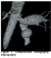
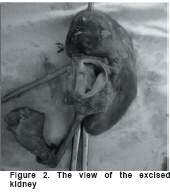
European Journal of General Medicine, Vol. 5, No. 4, 2008, pp. 251-253 Hypertension Due To Renal Artery Aneurysm Fuat Sar1, Cigdem Kutlu1, Yesim Kara1, Mehnur Turan1, Faik Cetin1, Omer Sarılar2, Rumeyza Kazancioglu3 Haseki Training and Research Hospital, Departments of Internal Medicine1, Urology2 and Nephrology3, Istanbul, Turkiye Code Number: gm08051 The most common pathogenetic lesion associated with renovascular hypertension is atherosclerosis. Next in etiologic incidence is arterial fibromuscular hyperplasia. The only other lesion common enough to warrant percentage classification is renal artery aneurysm. Here, we present a case with an aneurysm of the left renal artery following a gunshot wound and its resultant malign hypertension. Key words: Hypertension, renal artery aneurysm, renovascular. INTRODUCTION The most common pathogenetic lesion associated with renovascular hypertension is atherosclerosis. Next in etiologic incidence is arterial fibromuscular hyperplasia. The only other lesion common enough to warrant percentage classification is renal artery aneurysm (RAA) (1). Renal artery aneurysms are uncommon in vascular pathology with an incidence of 0.01-1%, though the first case was described by Rouppe in 1770 (2). Anatomically it has been recognized that an increase in blood pressure is based upon the formation of thrombosis within the RAA and that the degree of hypertension is related to the degree of occlusion and the diameter of the aneurysm (3). Traumatic aneurysm of the abdominal aorta secondary to penetrating injuries are rare (4, 5, 6). A few decades ago, the frequency of aneurysms was determined from post-mortem analysis and arteriographies performed for the assessment systemic hypertension. Accordingly Tcherdakoff found that 1.3% of patients undergoing arteriography had RAA (2). Here, we present a case with an aneurysm of the left renal artery following a gunshot wound and its resultant malignant hypertension resolving after nephrectomy. CASE A 16-year old female patient presented to our clinic with severe headache and fits. She had sustained hypertension for the last six months. In 2003, she underwent an intra-abdominal operation due to a self-inflicted gunshot injury to the abdomen. She did not smoke or use any illicit drug. Physical examination revealed an abdominal significant bruit below the median incision scar. Blood pressure was 210/120 mm Hg. The examination of the ocular fundus showed grade IV hypertensive retinopathy. Electro-cardiography had sinus rhythm and limited signs of left ventricular hypertrophy. Laboratory data were as follows: creatinine 106.4 mmol/L, sodium 136 mmol/L, potassium 4.3 mmol/L, calcium 2.6 mmol/L, renin 68.06 ng/(L.s). Computerized tomography angiography showed a distinct aneurismal dilatation in the proximal left renal artery (approximately 20 mm diameter) and accessory artery aneurysm (Figure 1). Scintigraphy revealed normal function of the right kidney while non functional kidney was detected on the left. Renovascular hypertension due to RAA was diagnosed. All other causes of RAA was discarded. Therefore; abdominal trauma was detected as the possible cause. She was referred to the surgery unit and total left nephrectomy was performed. The postoperative course was uneventful and her blood pressure decreased to 120/80 mm Hg. Since then the patient remained normotensive without any medication. DISCUSSION Renovascular hypertension is the most common cause of surgically correctable hypertension (1). Its cause in most younger individuals is fibromuscular dysplasia (about 40%), and the remainder of renal vascular disease is due to atherosclerotic stenoses of the proximal renal arteries (about 25%). Medial degeneration, trauma, injury are other potential causes (4). Arterial aneurysms have been described in the aorta and several major arteries including the renal arteries. They were associated with connective tissue diseases, Kawasaki disease, Takayasu disease, cystinosis, sepsis and fibromuscular dysplasia as well as neurofibromatosis and tuberous sclerosis (7). In our patient, all of these rare diseases were excluded based on clinical, laboratory and imaging findings. The incidence of RAA has increased in recent years, with the rising number of arteriographic explorations carried out for the study of systemic hypertension (2, 3). Many patients with renal artery aneurysm are asymptomatic and diagnosed as an incidental finding on CT scan or angiography performed for another purpose. Some patients have abdominal pain, palpable abdominal mass, subcostal or flank pain and hematuria. Almost 90% of symptomatic patients have systemic hypertension (2, 4). Our patient had also hypertension and abdominal bruit. Clinical characteristics such as age, sex and history play a little role in traumatic renovascular hypertension, but the sudden onset and rapid progression of hypertension in a young previously healthy individual who has suffered an abdominal trauma should provide an appropriate index of suspicion (1). Our patient had an operation due to gun shot wound of the abdomen. Direct arterial trauma is usually manifested by either laceration, transaction, contusion, or spasm. Laceration includes incomplete disruption of one or more layers of the wall (5). Evans and Moggs suggested that deceleration injury to the left renal artery is more common due to the comparative hypermobility of the left kidney as compared with the right (6, 8). In our patient, renal artery aneurysm was at the left kidney. The phenomenon of cured or improved hypertension in patients with RAA but without renal artery stenosis is hard to explain because the mechanism that might cause hypertension is difficult to show. Macroembolism or microembolism from the RAA into the renal parenchyma has been described, which can cause hypertension even without additional stenotic lesions of the renal artery. It is also possible that the RAA induces hemodynamic changes in terms of resistance, impedance, or shear stress at the vessel wall caused by turbulence, which may play a still unknown role (9). Aneurysms may rapidly develop to a large size and may potentially rupture; hence it represents a serious threat (2). Furthermore, although an effective antihypertensive drug treatment has been introduced in the recent years, hypertension still means an indication for the operative treatment of RAA (9). In the literature, the indication of RAA repair with regard to the prevention of aneurysm rupture is almost exclusively discussed in relation to aneurysm size. Although there is no clear correlation between maximal aneurysm diameter and the risk of rupture, on the basis of clinical experience, the treatment of RAAs with a diameter of 1.5 - 2 cm is generally recommended (9). The size of our patient’s aneurysm was 2 cm and the patient was transferred to the surgery unit. The standard surgical approach for renal artery aneurysms is excision with interposition grafting; if applicable. Infrequently, the aneurysm extends into the branch vessels and ex vivo reconstruction may be required. Our patient had total nephrectomy since there was no kidney function. The pathological examination of the excised kidney revealed subcapsular and intraparenchymal bleeding, double ureters and increased wall thickness (Figure 2). Renal artery aneurysm associated with renovascular hypertension due to abdominal trauma is rare. We emphasize the importance of thorough exploration of retroperitoneum following penetrating trauma in order to prevent future, sustained renal artery damage. REFERENCES
Copyright 2008 - European Journal of General Medicine The following images related to this document are available:Photo images[gm08051f2.jpg] [gm08051f1.jpg] |
| |||||||||
