 |
| About Bioline | All Journals | Testimonials | Membership | News |
|
||||||
|
||||||
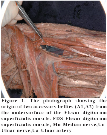
European Journal of General Medicine, Vol. 6, No. 1, 2009, pp. 57-59 Brief report Rare Origin of Two Accessory Bellies from the Undersurface of the Flexor Digitorum Superficialis Muscle Vasavi Rakesh G1, Bhagath Kumar Potu2, Raghu Jetti3, Venkata Ramana Vollala3, Thejodhar P4 Manipal University, KMC International Center, Department of Anatomy1, Kasturba Medical College, Department of Anatomy2, Melaka Manipal Medical College, Department of Anatomy3, Manipal, Karnataka, India, St. Matthew’s University School of Medicine, Department of Anatomical Sciences, Grand Cayman, Cayman Islands, British West Indies Code Number: gm09014 Proper knowledge of muscular variations is essential not only for anatomists but also for surgeon’s .Accessory bellies and the tendons of the muscles are surgically noteworthy. Such variant structures can lead to error in both diagnosis and treatment. Forearm flexors are known to exhibit such variations. Some of the variations are, the accessory heads of the deep flexors of the forearm (Gantzer’s muscles) have been described as 2 different small bellies which insert either into Flexor pollicis longus (FPL) or Flexor digitorum profundus (FDP). In 1813 Gantzer described 2 accessory muscles in the human forearm which bear his name (14,26) and these have subsequently been reported with variable attachments (6,11,12,14-16,19,24-26) .But there are no previous reports which have mentioned the existence of two accessory bellies arising from the undersurface of the flexor digitorum superficialis (FDS) and inserting into two deep flexors of the forearm as in the present case. In the routine dissection of right upper limb of 52 years old male cadaver in Department of Anatomy, K.M.C, Manipal, we observed a case of the flexor digitorum superficialis muscle giving two accessory bellies (A1,A2), which took their origin from the under surface of flexor digitorum superficialis just digital to the origin of this muscle from medial condyle . On further dissection we have noticed that, the accessory belly (A1) was running downwards to the medial aspect of the tendon of flexor pollicis longus for its insertion. The insertion was seen at the junction between proximal 1/3rd and middle 1/3rd of forearm; where as the other belly (A2) was running towards the lateral aspect of the tendon for the middle finger of flexor digitorum profundus for its insertion. The insertion of this belly was seen deep to the flexor retinaculum. The following are appropriate sizes of parts described. A1: Muscle length 5.9 cm, Tendon length 3.2 cm, Muscle width 0.6 cm, Tendon width 0.2 cm A2: Muscle length 7.2 cm, Tendon length 16.7 cm, Muscle width 0.8 cm, Tendon width 0.3 cm. The nerve supply for both muscle bellies came from the median nerve (Figure 1). Although many rare anatomical variations of FDS muscle were reported in the past, most of them appear to have no clinical significance. In recent times the variants have come to the notice because of their relationship with clinical problems requiring surgery (7). The FDS has been used as a motor for a wide variety of tendon transfer operations in the hand. The accessory heads of the deep flexors of the forearm (Gantzer’s muscles) have been described as two different small bellies which insert either into FPL or FDP. There are previous reports which have mentioned the existence of one accessory muscle which arose from the undersurface of the flexor digitorum superficialis and inserted into both FPL and FDP (10). Anomalies of the flexor digitorum Superficialis have been reported (1-4,8-10,14,21,23). The incidence of the accessory head of the FPL has been reported to range from 39±2% (26) to 73±7% (16) and that for the accessory head of the FDP from 2±9% (16) to 35±2 % (11). These accessory muscles have been observed to arise from the coronoid process, the medial epicondyle via fibres of the flexor digitorum superficialis, or a combination of the two (6, 11, 14-17, 22, 26). The accessory head of FPL has been observed anterior (16) or posterior to the anterior interosseous nerve (5). The accessory bellies described here are anterior to both the anterior interosseous nerve and vessels. The flexor muscles of the forearm develop from the flexor mass which subsequently divides into two layers, superficial and deep. The deep layer gives rise to the flexor digitorum superficialis, FDP and FPL (13). The existence of accessory muscles which connect the flexor muscles could be explained by the incomplete cleavage of the flexor mass during development. Apart from its anatomical interest, the accessory heads of the flexor muscles have been implicated in the anterior interosseous syndrome (5, 20) or as a cause of restricted movement of the FDP and FPL that can result in burning pain in the lower third of the forearm via a muscle-tendon shearing action (18). We conclude that such anomalous muscle bellies should be kept in mind while approaching the forearm for FDS tendon transfer and other surgical procedures around it. Acknowledgements We would like to thank Dr. Narga Nair, Prof & Head of Anatomy, K.M.C, Manipal for her support. REFERENCES
Copyright 2009 - European Journal of General Medicine The following images related to this document are available:Photo images[gm09014f2.jpg] [gm09014f1.jpg] |
| |||||||||
