 |
| About Bioline | All Journals | Testimonials | Membership | News |
|
||||||
|
||||||
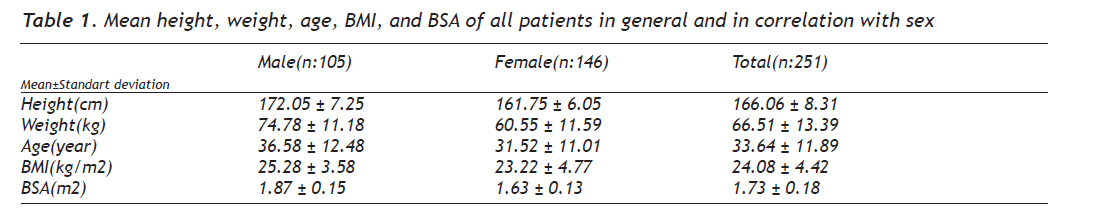
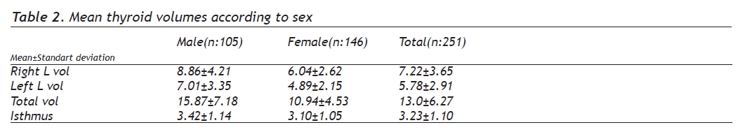
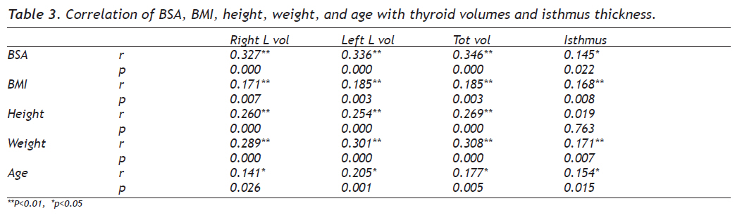
European Journal of General Medicine, Vol. 7, No. 2, April-June, 2010, pp. 125-129 Original Article Determination of Thyroid Volume and Its Relation with Isthmus Thickness Troid Volümünün Saptanması ve İstmus Kalınlığı ile İlişkisiServet Şeker¹, İsmet Taş2 Ankara Atatürk Education and Investigation Hospital, Department of Radiology¹, Van and Dr Muhittin Ülker Emergency and Traumatolgy Education and Investigation Hospital, Department of Radiology², Ankara, Turkey Correspondence: Dr Servet Şeker İstanbul Hastanesi Van, Turkey. Phone: 90 505 77 35 611 E-mail: mseker2@hotmail.com Received: 12.05.2009 Accepted: 14.08.2009 Code Number: gm10024 ABSTRACT Aim: Thyroid is a suitable organ for the investigation with ultrasonography due to its very superficial localization in body. It is also useful in frequent controls of the organ. The thyroid volume is variable among countries and its value in our country was not clearly demonstrated. In this study we aimed to determine the thyroid volume and its relation with isthmus thickness in Turkey. Key words: Ultrasonography, thyroid, anatomy, volume Amaç: Tiroid bezi yüzeyel yerleşimi nedeniyle ultrasonografik incelemeye elverişli bir organdır. Tiroid bezi hastalıklarında sık kontrollere ihtiyaç duyulması da ultrasonografinin önemini artırmaktadır. Tiroid bezi volümü ülkeden ülkeye belirgin değişiklikler göstermekte olup bu konuda ülkemizde net kabul edilmiş değerler mevcut değildir. Biz bu çalışmada Türkiye’de tiroid bezi volümünü ve istmus kalınlığı ile ilişkisini araştırmayı amaçladık. Anahtar kelimeler: Ultrasonografi, troid, anatomi, volüm INTRODUCTION Thyroid is a suitable organ for the investigation with ultrasonography due to its superficial localization in body. More over this modality is the sole modality that must be preferred in diffuse thyroid diseases (1). It is also useful in frequent controls of the organ. The thyroid volume is variable among countries and its value in our country was not clearly demonstrated. In this study we aimed to determine the thyroid volume and its relation with isthmus thickness in Turkey. MATERIALS AND METHODS A total of 251 volunteers without any complaint or physical examination finding regarding the thyroid disease were included in the study in which 105 man and 146 women, age range between 15 and 78. Patients with prior thyroid disease history or the patients in whom nodule, heterogeneity or agenesis were discovered in the USG examination were excluded from the study. EUB-6000 Hitachi USG device with 10 Mhz lineer probe in “Dr.Muhittin Ülker Emergency and Traumatology Hospital” Radiology Department was used. No prior preparation was done and studies were conducted by one dedicated operator. Thyroid gland size measurements were done once but in case of imaging problems due to body composition differences (i.e. short neck, kyphosis etc) multiple measurements were done to provide accurate results. Craniocaudal length, transverse and mediolateral length (width), antero posterior length for each lobe and isthmus thickness were measured and recorded. The volume of each thyroid lobe was calculated with ellipsoid formula: (Volume (ml) = Length (cm) x Width (cm) x Thickness (cm) x 1/6 Л ). Total volume was obtained as the sum of two thyroid lobes. The isthmus was not included into the sum (2-5). Body mass index (BMI) (kg/m2) was calculated as= weight / height2 Body surface area (BSA) (m2) was calculated as= weight0,425 x height0,725 x 71,84 x 10-4 (3). In statistical analysis, normal distribution was studied with Kolmogorov-Smirnov test. The correlation between sex and thyroid volumes, right and left lobe volumes, total volume and isthmus thickness were evaluated with Independent-samples T test. The differences of the variables in each group (e.g. right thyroid lobe volume and left thyroid lobe volume in males) were evaluated with Paired-Samples T test. The correlation among other variables was evaluated with Pearson correlation test. p< 0.05 was accepted statistically significant. RESULTS In our study, 105 men and 146 women were included, and the mean age was 36.58 and 31.52, respectively. The mean age for all patients was 33.64. In Table 1, mean height, weight, age, BMI, and BSA of all patients in general and in correlation with sex are summarized. In Table 2, mean thyroid volumes are summarized according to sex. All the thyroid volumes are bigger in men than in women. In Table 3, the correlation of 5 parameters (BSA, BMI, height, weight, and age) with thyroid volumes and isthmus thickness is summarized. A significant correlation was found when BSA, BMI, weight, and age were correlated with thyroid volumes and isthmus thickness. There was also a significant correlation between height and thyroid volumes. Significant correlation was found when BSA and weight were correlated with all thyroid volumes. There was a correlation between height and craniocaudal thyroid volumes in men. Mean thyroid volumes and mean isthmus thickness is summarized in graphic 1. Mean thyroid dimensions according to sex are shown in graphic 2. In our study the patients were subdivided into 6 groups according to age. The mean thyroid volumes were found to be 10 ml in 15-24 age group (52 patients), 13 ml in 25-34 age group (104 patients), 14 ml in 35-44 age group (63 patients), 16 ml in 45-54 age group (15 patients), 17 ml in 55-64 age group (9 patients), and 12 ml in over- 65 age group (9 patients). Owing to these results, there was a positive correlation between age and total thyroid volumes up to 65 years. A negative correlation was found after 65 years, which we suppose not confident due to the low number of patients in this group. DISCUSSION Thyroid gland lies anterior to the neck, beside the laryngotracheal axis, at the subhyaloid portion. The adult thyroid gland weights 20-30 grams and is butterfly-shaped. Enlargement of the thyroid gland is due to several factors, such as hormonal or immunogical stimulation, inflammatory, proliferative, infiltrative, or metabolic disorders (6). A normal thyroid gland volume does not exclude the diagnosis of nodulary goitre (7). Estimating the volume of a thyroid gland via palpation has low sensitivity and specifity. In this context thyroid ultrasonography is a reliable method in calculating the volume of a thyroid gland (8). The high resolution property of ultrasonography gives the opportunity of evaluating the morphology, dimensions, and paranchimal structure of a thyroid gland adequately. Additionally, ultrasonography is the basic method to calculate the volume of the gland. Scintigraphy supplies data about the local functions of the thyroid gland, which cannot be obtained via the rest of the methods. Computed tomography or magnetic resonance imaging has limited value in the diagnoses of the thyroid gland diseases (9). We investigated any relation between the dimension, volume, or the thickness of isthmus of the thyroid gland and length, age, gender, BMI, or BSA. Relations between the parameters mentioned above and volume or dimensions of the thyroid gland has been investigated by many investigators, who have reported various results. Moreover, the volume of the thyroid gland varies fron nation to nation. Berghout et al.10 reported a mean adult thyroid gland volume of 10.7±4.6 ml in healthy adults in Netherlands in 1987. Berghout et al. (10) also reported that the mean male thyroid gland volume (12.7±4.4) was higher than the mean female thyroid gland volume (8.7±3.9). Hegedus et al11. reported a mean adult thyroid gland volume of 18.6±4.5 ml in 271 healthy adults in 1983. Hegedus et al (11). reported a significant difference between the mean male thyroid gland volume (19.6±4.7 ml) and the mean female thyroid gland volume (17.5±4.2) and concluded adult thyroid gland volume was correlated with body weight and age. Ueda reported from Japan that thyroid volume was correlated with age, height, weight and BSA. He could not find any difference between man and women (12). Nygaard et al from Denmark noted that thyroid volumes were 12 ml (4-29 ml), 18 ml (5-47 ml), 18 ml (7-64 ml) and 18 ml (9-51 ml for 4 age groups lines as 15, 30, 45 and 60 respectively in 391 women from Denmark (13). Barrere et al from France reported that thyroid volume was positively correlated with age, height, weight and BSA in both gender and negatively correlated with age in women. In the same study also intresting is the finding that thyroid volumes were bigger in smokers and ex smokers, also smaller in oral contraceptive users (14). Oberhofer et al reported the mean thyroid volume as 13.35 ml (man 14.94 m, women 12.09 ml in 500 healthy adults at 1989in Austria (15). Gomez et al reported the thyroid volume as 9.8±4.6 ml form an and 6.5±2 ml for women (16). In our study these volumes were 15.87±7.18 ml and 10.94±4.53 ml respectively (mean 13±6 ml) (p=0.000). Mean isthmus thickness was 3.42±1.14 mm in man and 3.10±1.05 mmin women (mean 3.23±1.10 mm)(p=0.021). When right and left lobes compared in man right lobe was 8.86±4.21 ml,in women 6.04±2.62 ml,left lobe in man 7.01±3.35 ml and in women 4.89±2.15 ml (p<0.05). Right lobe is bigger in both sexes. Reported normal thyroid gland dimensions were variable; in one study the thickness was 15-20 mm, width 20-25 mm and length was 30-50 mm. In another study length was 40- 60 mm, thickness 13-18 mm and isthmus was 3-8 mm (17). In conclusion in our study longitudinal length of the thyroid was longer in man and right lobe is longer than the left one. This is true for a-p length of the gland and transverse length. When both sexes studied together BSA was correlated with total thyroid volume, right lobe volume, left lobe volume, all dimensions of the thyroid and isthmus thickness. When man and women separately studied, only left lobe transverse dimension was correlated with BSA. Also BMI was correlated with total thyroid volume, right lobe volume, left lobe volume and isthmus thickness when both sexes studied together. This not valid for man when separated, also when women group considered BMI was correlated only with total thyroid volume, right lobe volume and left lobe volume. BMI was correlated with right and left lobe a-p and transverse dimensions, no correlation was observed with craniocaudal dimensions. When sexes separated no correlation was present. In general, age was correlated with a-p dimensions of the both lobes, right lobe volume, left lobe volume, total volume and isthmus thickness. In this case the correlation was different from the general in women. REFERENCES
Copyright 2010 - European Journal of General Medicine The following images related to this document are available:Photo images[gm10024t2.jpg] [gm10024t1.jpg] [gm10024f2.jpg] [gm10024t3.jpg] [gm10024f1.jpg] |
| |||||||||
